Hello! Welcome to your fourth lesson in the "Female Reproductive Anatomy and Physiology" module.
In our last lesson, we examined the uterus and the dynamic changes of the endometrium. We learned that after ovulation, the endometrium enters a "secretory phase," where it becomes thick, vascular, and rich in nutrients, all to prepare a welcoming environment for a potential embryo. This change, we noted, is driven by the hormone progesterone. Today, we will uncover the source of that critical hormone.
This lesson addresses the learning outcome: Describe the formation, function, and regression of the corpus luteum. We will explore the remarkable transformation of the follicle after it has released its egg, turning into a temporary but powerful endocrine gland. Understanding the life cycle of the corpus luteum is the key to linking the events in the ovary (the ovarian cycle) with the changes in the uterus (the uterine cycle).
1. Formation of the Corpus Luteum: A Phoenix from the Ashes
After the dramatic event of ovulation, where the mature Graafian follicle ruptures to release the oocyte, the story of the follicle is not over. The remaining structure collapses and undergoes a rapid transformation.
To see this process, let's start with a short video that explains what happens immediately following ovulation.
Menstrual Cycle Phases | Female Reproductive System | Follicular Luteal Proliferative Secretory
The video 'Menstrual Cycle Phases' by RegisteredNurseRN provides a very clear explanation of how the remnant of the follicle becomes the corpus luteum.
Please watch the segment from 06:12 to 07:07. Focus on how the LH surge not only triggers ovulation but also initiates the transformation of the ruptured follicle into this new structure, the corpus luteum.
As the video highlighted, Luteinizing Hormone (LH) is the key trigger. The process it initiates is called luteinization. Here’s a more detailed breakdown:
- The remaining follicular cells—the granulosa and theca cells—change dramatically.
- They enlarge and begin to accumulate cholesterol and a yellow pigment called lutein. This pigment gives the structure its name: corpus luteum, which is Latin for "yellow body."
- The basement membrane that once separated the granulosa and theca cells breaks down, allowing blood vessels to rapidly grow into the center of the structure. This intense vascularization, or angiogenesis, is essential because the corpus luteum has one of the highest rates of blood flow per unit of tissue in the entire body, reflecting its high metabolic and hormonal activity.
For a more detailed textual description of this transformation, please read the following resource.
The Normal Menstrual Cycle and the Control of Ovulation
The article 'The Normal Menstrual Cycle and the Control of Ovulation' from Endotext gives a concise, high-level description of the luteinization process.
Please read the first paragraph under the heading 'LUTEAL PHASE'. It clearly describes how luteinized granulosa and theca-lutein cells combine to form the corpus luteum and highlights its vascularization.
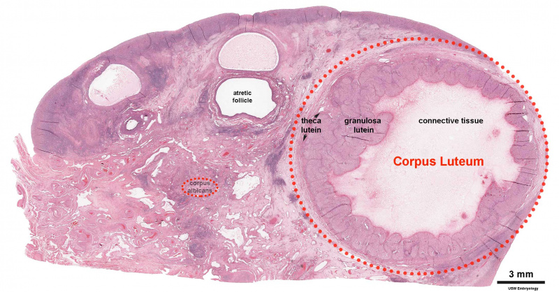
At a microscopic level, we can see the resulting structure clearly.
2. The Function of the Corpus Luteum: A Temporary Hormone Factory
The primary function of the corpus luteum is to act as a temporary endocrine organ, and its hormonal output is crucial for the establishment and maintenance of early pregnancy. It secretes several key hormones, which are reflected in the hormonal changes of the menstrual cycle.
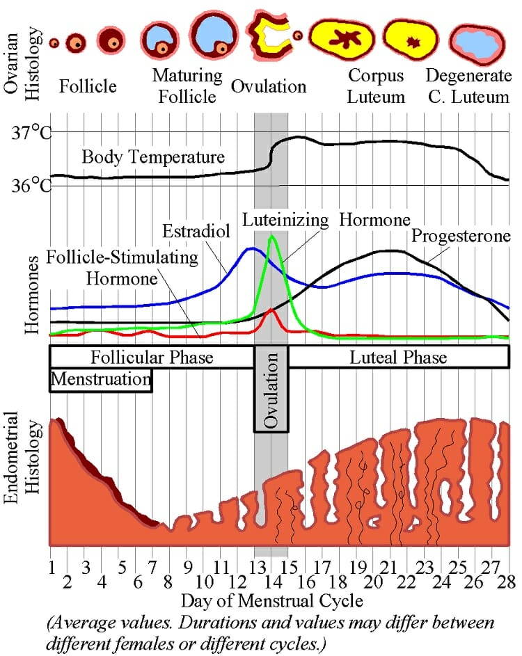
Let's look at the hormones and their functions:
-
Progesterone: This is the dominant and most important hormone of the luteal phase. Its name is a clue to its function: "pro-gestation."
- Action on the Uterus: It acts on the estrogen-primed endometrium, causing it to enter the secretory phase we discussed in the last lesson. The uterine glands become coiled and secrete glycogen, and the spiral arteries grow, making the lining thick and receptive to implantation.
- Action on the HPO Axis: Progesterone exerts strong negative feedback on the hypothalamus and pituitary, suppressing the release of GnRH, LH, and FSH. This prevents the development of new follicles during the luteal phase.
-
Estradiol (Estrogen): The corpus luteum also secretes estrogen. This estrogen works synergistically with progesterone to help maintain the endometrial lining.
-
Inhibin A: This hormone is also produced by the corpus luteum. It specifically acts on the anterior pituitary to inhibit the secretion of FSH, further ensuring that no new follicular recruitment occurs.
This hormonal output is not a steady stream but is released in pulses, driven by the pulsatile secretion of LH from the pituitary gland that continues throughout the luteal phase.
Test your understanding!
During the luteal phase, the corpus luteum secretes high levels of progesterone and inhibin A. What is the combined effect of these two hormones on the pituitary gland, and why is this effect biologically important at this stage of the cycle?
Show answer
The combined effect is a powerful suppression of both FSH and LH secretion from the pituitary gland. Progesterone primarily suppresses LH (and GnRH), while inhibin A specifically suppresses FSH.
This is biologically crucial because it prevents the recruitment and development of a new cohort of ovarian follicles. If new follicles were to develop and ovulate while the uterus is already preparing for a potential pregnancy from the current cycle, it would lead to hormonal chaos and disrupt the chances of a successful pregnancy. This hormonal "quieting" ensures that the body's resources are focused on supporting the potential implantation and early development of an embryo.
3. The Two Fates of the Corpus Luteum: Regression or Rescue
The corpus luteum has a pre-programmed, finite lifespan of approximately 14 days. Its ultimate fate is binary: it will either degenerate if no pregnancy occurs, or it will be "rescued" and continue to function if pregnancy is established.
The following video provides an excellent summary of these two pathways.
Menstrual Cycle Phases | Female Reproductive System | Follicular Luteal Proliferative Secretory
Let's return to the 'Menstrual Cycle Phases' video by RegisteredNurseRN. It does a great job explaining the two different fates of the corpus luteum.
Please watch from 11:59 to 15:19. Pay close attention to the two scenarios presented: What happens when there is no fertilization (disintegration). What happens when fertilization occurs (the role of hCG).
Let's explore these two fates in more detail.
Path 1: No Pregnancy → Regression (Luteolysis)
If the oocyte is not fertilized and implantation does not occur, the corpus luteum begins to self-destruct around 9-11 days after ovulation. This process of programmed degeneration is called luteolysis.
- Mechanism: While the exact mechanism in humans is complex, it is understood to involve local factors within the ovary, such as prostaglandins (specifically Prostaglandin F2α), which inhibit steroid production and promote cell death (apoptosis) within the corpus luteum.
- Transformation: As the luteal cells die, the corpus luteum shrinks and is replaced by avascular scar tissue. This pale, scar-like remnant is called the corpus albicans (Latin for "white body"). You can see a corpus albicans in the histology image we viewed earlier (resource LINK).
- Consequences: The degeneration of the corpus luteum leads to a sharp decline in the blood levels of progesterone and estrogen. This hormone withdrawal has two immediate and critical effects:
- The endometrial lining, no longer supported by progesterone, breaks down and is shed, resulting in menstruation.
- The negative feedback on the hypothalamus and pituitary is removed. This allows FSH levels to rise again, initiating the recruitment of a new cohort of follicles for the next cycle.
Path 2: Pregnancy → Rescue by hCG
If fertilization and implantation do occur, the developing embryo must signal its presence to prevent the self-destruction of the corpus luteum.
- The Rescue Signal: About 8-9 days after fertilization, the outer layer of the embryo (the syncytiotrophoblast) begins to secrete a hormone called human Chorionic Gonadotropin (hCG). This is the hormone detected in pregnancy tests.
- Mechanism of Action: hCG is structurally very similar to LH and functions as a potent LH analogue. It binds to the LH receptors on the cells of the corpus luteum, overriding the luteolytic process and stimulating the corpus luteum to continue producing progesterone.
- The Corpus Luteum of Pregnancy: This "rescued" structure is now called the corpus luteum graviditatis. It is essential for survival of the early pregnancy, as its progesterone output maintains the endometrium and prevents menstruation. It continues this vital function until the placenta is sufficiently developed to take over progesterone production, which typically occurs around the 8th to 12th week of gestation. After this point, the corpus luteum graviditatis finally regresses.
Conclusion
Today, we've followed the full life story of the corpus luteum, a transient but indispensable player in female reproductive physiology.
Key Takeaways:
- The corpus luteum forms from the remnants of the ovulated follicle in a process called luteinization, driven by the LH surge.
- It functions as a temporary endocrine gland, with its primary role being the secretion of progesterone, which makes the uterus receptive to pregnancy and suppresses further follicular development.
- If pregnancy does not occur, the corpus luteum regresses after about 14 days into a scar tissue called the corpus albicans. The resulting drop in progesterone triggers menstruation.
- If pregnancy occurs, the embryo produces hCG, which "rescues" the corpus luteum, ensuring continued progesterone production to sustain the early pregnancy.
Preview of the Next Lesson:
This lesson concludes our module on the fundamental anatomy and physiology of the female reproductive system. We've covered the ovary, folliculogenesis, the uterus, and the corpus luteum. In our next lesson, we will begin a new module, "The Hypothalamic-Pituitary-Ovarian (HPO) Axis." We will take a step back to look at the master controllers of this entire system: the brain and pituitary gland, and explore the intricate hormonal conversations that orchestrate the entire menstrual cycle.