Hello! Welcome to your third lesson in the "Female Reproductive Anatomy and Physiology" module.
In our last lesson, we traced the long and fascinating journey of folliculogenesis, from a dormant primordial follicle to a mature, hormone-producing Graafian follicle ready for ovulation. We saw how this process is essential for producing a viable egg.
Today, we shift our focus from the site of egg production (the ovary) to the site of potential pregnancy. We will address the learning outcome: Describe the structural and functional anatomy of the uterus and endometrium. The uterus is a remarkable organ that must prepare itself each month to receive and nourish a fertilized egg. Understanding its structure and the dynamic changes it undergoes is fundamental to understanding the menstrual cycle and, later, the menstrual irregularities that are a hallmark of PCOS.
1. Gross Anatomy of the Uterus
Let's begin with the overall structure and location of the uterus. It is a hollow, pear-shaped muscular organ situated in the female pelvis, typically positioned between the urinary bladder and the rectum.
To get a clear picture of its major parts, size, and typical orientation, the following video provides an excellent overview.
Anatomy of Uterus - Parts and position, Blood supply, Nerve supply, Lymphatic drainage - Part 1
The video 'Anatomy of Uterus' from MedSchool Simplified offers a clear, concise introduction to the gross anatomy of the uterus.
Please watch from 00:42 to 04:29. As you watch, focus on identifying the four main anatomical regions and understanding the terminology for uterine position: Fundus: The dome-shaped top of the uterus. Corpus (Body): The main, central part. Isthmus: The narrow transition zone between the body and the cervix. Cervix: The lower, cylindrical part that projects into the vagina. Anteflexion and Anteversion: The typical forward tilt of the uterus.
As the video explained, the uterus is not just a static structure. Its size and the ratio of its parts change throughout a woman's life.
- Pre-puberty: The uterus is small, and the cervix is proportionally larger than the body.
- Reproductive Years: The uterine body grows significantly under hormonal influence, becoming much larger than the cervix.
- Post-menopause: With the decline in hormones, the uterus atrophies (shrinks), and the body becomes smaller again relative to the cervix.
These anatomical details form the foundation for understanding its function. Now, let's look inside the uterine wall.
2. The Uterine Wall: A Three-Layered Structure
The wall of the uterus is composed of three distinct layers, each with a specific function.

The three layers, from outermost to innermost, are:
- Perimetrium: The thin, outer serous layer. It is part of the peritoneum, the membrane that lines the abdominal cavity, and it covers the majority of the uterus.
- Myometrium: The thickest layer, composed of powerful smooth muscle fibers. This muscular wall gives the uterus its strength. Its primary functions are to expand dramatically during pregnancy to accommodate a growing fetus and to produce strong, coordinated contractions during labor and menstruation.
- Endometrium: The innermost mucosal lining. This is the most dynamic layer of the uterus and the one we will focus on most, as it is central to the menstrual cycle and pregnancy.
3. The Endometrium: The Functional Heart of the Uterus
The endometrium is not a single, uniform layer. It is divided into two distinct sub-layers with different functions and fates during the menstrual cycle.
- Stratum Functionale (Functional Layer): This is the superficial layer that lines the uterine cavity. It is this layer that proliferates and thickens each month under hormonal influence to prepare for a potential embryo implantation. If pregnancy does not occur, the entire functional layer is shed during menstruation.
- Stratum Basale (Basal Layer): This is the deeper, permanent layer that lies adjacent to the myometrium. It is not shed during menstruation. Its crucial function is to act as a regenerative source, from which a new functional layer grows after each period.
The endometrium also contains simple tubular glands, known as uterine glands, which extend from the surface down into the basal layer. These glands produce a glycogen-rich fluid that provides nourishment for an early embryo.
To understand how the functional layer can be shed while the basal layer remains intact, we need to examine their unique blood supply.
Fallopian tubes and uterus: Histology
The 'Fallopian tubes and uterus: Histology' video from Osmosis provides an outstanding visual explanation of the endometrial layers and their specialized blood supply.
Please watch from 04:37 to 07:41. Pay close attention to: The visual distinction between the functional layer and the basal layer. How the uterine blood supply branches into straight arteries and spiral arteries. Which layer each type of artery supplies and why this is so important.
As the video explained, the blood supply is key:
- Large arcuate arteries run within the myometrium.
- They give rise to radial arteries that penetrate towards the endometrium.
- These then divide:
- Straight arteries supply the permanent basal layer (stratum basale).
- Spiral arteries grow into the temporary functional layer (stratum functionale). These arteries are sensitive to hormones and are crucial for both building up the endometrium and initiating its breakdown during menstruation.
This dual blood supply is a brilliant piece of biological engineering, allowing for the monthly renewal of the uterine lining without compromising the organ's integrity.
Test your understanding!
A patient undergoes a surgical procedure called endometrial ablation, where the lining of the uterus is destroyed to treat heavy menstrual bleeding. In a successful procedure, the patient stops having periods permanently. Based on your knowledge of the endometrial layers, which layer must be destroyed to achieve this outcome, and why?
Show answer
To permanently stop menstruation, the stratum basale (basal layer) must be destroyed.
Reasoning: The stratum functionale is naturally shed every month. If only the functional layer were destroyed, the basal layer would simply regenerate a new functional layer in the next cycle, and periods would continue. The basal layer contains the stem cells responsible for rebuilding the endometrium. By destroying the stratum basale, the regenerative source is eliminated, and a new functional layer cannot be formed.
4. Functional Changes: The Uterine Cycle
The structure of the endometrium is not static; it undergoes a predictable cycle of changes each month, driven by the ovarian hormones estrogen and progesterone. This is known as the uterine (or menstrual) cycle.
Let's explore the three phases of this cycle. For a comprehensive understanding, we will use a combination of video and text.
Uterus: Anatomy, blood supply, histology, functions
The Kenhub article 'Uterus: Anatomy, blood supply, histology, functions' provides a good textual description of the phases.
Please read the section titled 'Function'. It clearly outlines the menstrual, proliferative, and secretory phases and the key events in each.
Now, let's visualize these changes.
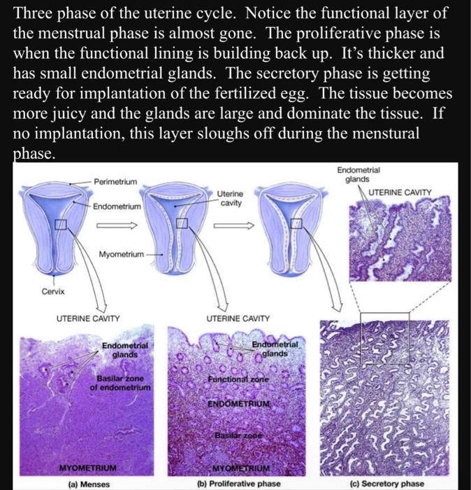
Here’s a summary of the phases, connecting them to the hormonal changes we will study later:
-
Menstrual Phase (Days 1-5):
- If no pregnancy occurs, ovarian hormone levels (estrogen and progesterone) drop.
- This causes the spiral arteries in the stratum functionale to constrict, cutting off blood supply.
- The functional layer becomes ischemic (lacks oxygen), dies, and is shed from the uterus, resulting in menstrual bleeding.
-
Proliferative Phase (Days 6-14):
- This phase is driven by estrogen, which is produced by the growing follicles in the ovary.
- Cells in the stratum basale multiply rapidly, regenerating a new stratum functionale.
- The endometrium thickens, and the uterine glands are relatively straight and narrow.
-
Secretory Phase (Days 15-28):
- This phase is driven by progesterone, which is produced by the corpus luteum in the ovary after ovulation.
- Progesterone causes the endometrium to become receptive to implantation.
- The uterine glands become coiled and "saw-toothed" in appearance and begin to secrete glycogen.
- Blood supply to the functional layer increases further. The uterus is now fully prepared for pregnancy.
If implantation does not occur, the cycle repeats as progesterone levels fall, triggering menstruation.
Conclusion
In this lesson, we have explored the anatomy of the uterus, from its macroscopic structure down to the microscopic and functional details of its inner lining, the endometrium.
Key Takeaways:
- The uterus is a muscular organ with three layers: the outer perimetrium, the powerful middle myometrium, and the dynamic inner endometrium.
- The endometrium is comprised of a permanent, regenerative stratum basale and a temporary stratum functionale that is shed during menstruation.
- A unique dual blood supply, with straight arteries for the basal layer and hormone-sensitive spiral arteries for the functional layer, facilitates this monthly cycle.
- The endometrium undergoes cyclical changes—the proliferative phase (estrogen-driven rebuilding) and the secretory phase (progesterone-driven preparation for implantation)—to prepare for pregnancy each month.
Preview of the Next Lesson:
We've now covered follicular development in the ovary and the preparation of the uterus. What connects these two events? The answer lies in what happens to the follicle after it ovulates. In our next lesson, "Describe the formation, function, and regression of the corpus luteum," we will examine this temporary endocrine structure that forms from the ruptured follicle. You'll learn how its production of progesterone is the critical signal that drives the secretory phase in the endometrium we just discussed, tying the ovarian and uterine cycles together.