Hello! Welcome to the final lesson in our module on Female Reproductive Anatomy and Physiology.
In our last session, we explored the fascinating life cycle of the corpus luteum, from its formation after ovulation to its critical role in producing progesterone. We saw how its fate—either regression or rescue by hCG—determines whether a menstrual cycle ends or a pregnancy is sustained. The regression of the corpus luteum and the subsequent fall in hormones is the event that resets the system, allowing a new cycle to begin.
Today's lesson addresses the learning outcome: Explain the concept of the ovarian reserve and age-related changes in ovarian function. We will zoom out to look at the bigger picture: the total supply of eggs a woman has throughout her life. This "ovarian reserve" is the fundamental resource from which each cycle's follicles are drawn. Understanding its finite nature and how it changes with age is essential for grasping the full context of female fertility and is a cornerstone for our future discussions on PCOS.
This lesson concludes our deep dive into the ovary itself. Having covered its structure, the process of folliculogenesis, and the corpus luteum, understanding the ovarian reserve will complete your foundational knowledge before we move on to explore its regulation by the brain in the next module.
1. The Ovarian Reserve: A Woman's Finite Follicular Pool
Unlike men, who produce new sperm throughout their lives, women are born with all the eggs (oocytes) they will ever have. These oocytes are housed within structures called primordial follicles. The total number of these follicles in the ovaries at any given time is known as the ovarian reserve.
This reserve is not static; it is established before birth and undergoes a continuous, irreversible decline throughout a woman's life.
To get a sense of the scale of this decline, please watch the first part of the following video.
The video 'Female Aging and Fertility' by Dr. Anu Kathiresan provides an excellent and concise overview of the decline in egg quantity over a woman's lifespan.
Please watch the segment from 01:00 to 01:45. Pay close attention to the specific numbers mentioned at different life stages: fetal life, birth, puberty, and menopause.
As the video explained, the numbers are dramatic:
- Peak in Fetal Life: A female fetus has the maximum number of oocytes at around 20 weeks of gestation, peaking at 6 to 7 million.
- At Birth: This number has already dropped significantly to 1 to 2 million.
- At Puberty: The reserve has further dwindled to about 300,000 to 400,000.
- At Menopause: Fewer than 1,000 follicles remain.

You might wonder where all these follicles go. Of the hundreds of thousands present at puberty, only about 400-500 will ever be ovulated. The vast majority are lost through a process of programmed cell death called atresia. This is a continuous process that happens regardless of whether a woman is pregnant, has regular cycles, or is using hormonal contraception.
For a more detailed explanation of this process, please read the following text.
Volume 5, Chapter 72. Age and Reproduction
The chapter 'Age and Reproduction' provides an in-depth look at the establishment of the follicular pool and the process of atresia.
Please read the section titled 'Ovarian Changes'. Focus on the paragraphs describing the initial formation of the oocyte pool during gestation and the subsequent massive depletion through follicular atresia before and after birth.
2. The Impact of Aging: A Decline in Both Quantity and Quality
As a woman ages, the decline in her ovarian reserve has two critical dimensions: the number of eggs (quantity) and their biological fitness (quality).
Decline in Quantity
As we've seen, the number of follicles steadily decreases over time. However, this decline is not linear. Research shows that the rate of follicular loss accelerates around the age of 37, when approximately 25,000 follicles remain. This accelerated depletion is what precedes the transition into perimenopause and eventually menopause.
Decline in Quality
Perhaps even more important than the decrease in quantity is the decline in oocyte quality. "Quality" primarily refers to the genetic integrity of the egg, specifically whether it has the correct number of chromosomes.
As oocytes age along with the woman, they become more prone to errors during the final stages of meiosis (cell division), which occurs just before ovulation. This leads to a higher incidence of aneuploidy—oocytes with an abnormal number of chromosomes.
An aneuploid egg, if fertilized, is much less likely to result in a healthy pregnancy. This is the primary biological reason why advancing maternal age is associated with:
- Decreased fertility (lower chance of conception per month).
- Increased rates of miscarriage.
- Higher risk of chromosomal disorders in offspring (like Down syndrome).
The next video segment explains this concept clearly.
Let's return to the 'Female Aging and Fertility' video to understand the concept of egg quality.
Watch the segment from 01:45 to 02:23. The video explains how aging affects the process of chromosome division in the egg.
An important question is whether poor reserve is just about having fewer eggs, or if it also implies the remaining eggs are of lower quality. While age is the main driver of declining quality, evidence suggests that women with a lower-than-expected ovarian reserve for their age also tend to have a higher proportion of abnormal eggs. The following video explores this relationship.
Does low AMH / low ovarian reserve mean worse eggs? Or just fewer?
The video 'Does low AMH / low ovarian reserve mean worse eggs?' delves into the scientific evidence linking egg quantity and quality.
Watch the segment from 01:59 to 03:52. This part explains how researchers have controlled for age to show that diminished ovarian reserve itself is associated with a higher rate of chromosomally abnormal embryos.
3. Clinical Assessment of Ovarian Reserve
Since we cannot directly count the microscopic primordial follicles, clinicians use indirect markers to estimate a woman's ovarian reserve. These "ovarian reserve tests" (ORTs) help predict a woman's reproductive potential, particularly in the context of fertility treatments.
The most common tests fall into two categories: blood tests (biochemical markers) and ultrasound imaging (biophysical markers).
The following reading provides a clear and concise summary of the most important tests used today.
An Update on Ovarian Aging and Ovarian Reserve Tests
The review article 'An Update on Ovarian Aging and Ovarian Reserve Tests' provides an excellent overview of the modern tools used to assess ovarian reserve.
Please read the section 'Ovarian reserve tests (ORTs)'. Focus on understanding the principles behind Anti-Mullerian Hormone (AMH), Antral Follicle Count (AFC), and Basal Follicle Stimulating Hormone (FSH).
Let's summarize the key tests mentioned in the article:
-
Anti-Müllerian Hormone (AMH):
- What it is: A hormone produced by the granulosa cells of small, growing (pre-antral and small antral) follicles.
- What it tells us: Since AMH is produced by the pool of developing follicles, its level in the blood correlates well with the size of the remaining primordial follicle pool. A higher AMH suggests a larger reserve, while a low AMH suggests a diminished reserve. It is considered one of the most reliable markers.
-
Antral Follicle Count (AFC):
- What it is: A direct count of the small (2-10 mm) follicles visible in both ovaries using a transvaginal ultrasound during the early part of the menstrual cycle.
- What it tells us: Like AMH, the number of visible antral follicles serves as a proxy for the size of the deeper, microscopic reserve. A low AFC is indicative of a diminished reserve.
-
Basal Follicle-Stimulating Hormone (FSH):
- What it is: A blood test measuring the level of FSH on day 2 or 3 of the menstrual cycle.
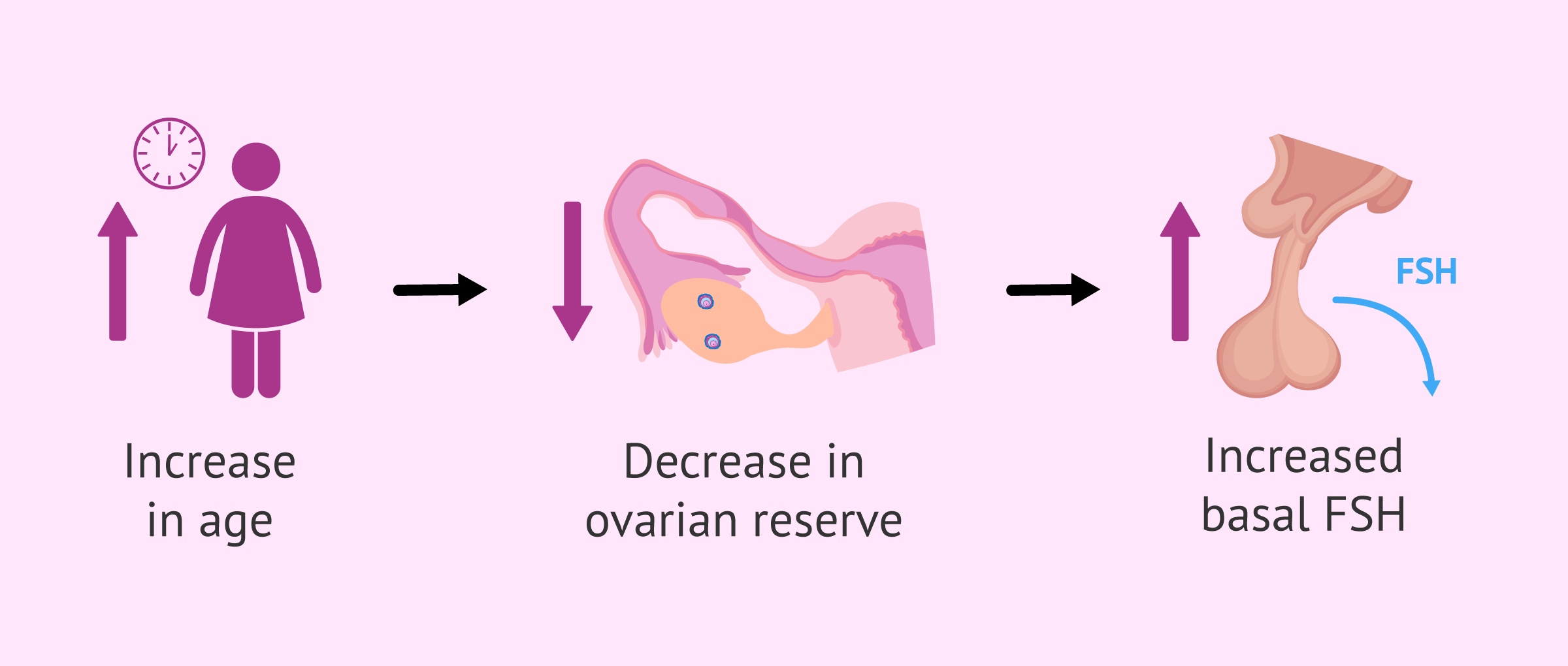
- What it tells us: This test is an indirect measure of the feedback loop between the ovaries and the pituitary gland. As the ovarian reserve declines, the ovaries produce less inhibin B (a hormone that suppresses FSH). To compensate, the pituitary gland must produce more FSH to stimulate the remaining follicles to grow. Therefore, an elevated basal FSH level is a sign of diminished ovarian reserve. The pituitary has to "shout" louder to get a response from the aging ovary.
Test your understanding!
A 39-year-old woman with regular menstrual cycles undergoes fertility testing. Her Day 3 FSH level is found to be consistently elevated. Based on what you've learned in this lesson, what does this finding suggest about her ovarian reserve, and what is the physiological reason for this elevation?
Show answer
The elevated Day 3 FSH level suggests she has a diminished ovarian reserve.
The physiological reason is rooted in the negative feedback loop of the HPO axis. Her smaller pool of remaining follicles produces lower levels of hormones like inhibin B. Inhibin B's job is to suppress FSH secretion from the pituitary. With less inhibin B, the pituitary is less inhibited and therefore releases more FSH in an attempt to adequately stimulate the fewer, less responsive follicles in her ovaries. The elevated FSH is a compensatory response to declining ovarian function.
Conclusion
This lesson completes our journey through the fundamental physiology of the ovary. We've established that the ovary's function is dictated by a finite resource that declines predictably with age.
Key Takeaways:
- Ovarian reserve refers to the limited pool of follicles a woman is born with, which depletes continuously through a process called atresia.
- Advancing age leads to a decline in both the quantity (number) and quality (genetic health) of the remaining oocytes. This decline accelerates in a woman's late 30s.
- This age-related decline is the primary cause of reduced fertility and increased miscarriage rates in older women.
- Clinicians estimate ovarian reserve using tests like AMH, Antral Follicle Count (AFC), and basal FSH levels, which provide an indirect measure of reproductive potential.
Preview of the Next Lesson:
We now have a solid understanding of the events within the ovary: how follicles grow, how one is selected for ovulation, what happens to its remnant (the corpus luteum), and how the total supply of follicles changes over a lifetime.
With this foundation in place, we are ready to move to Module 3: The Hypothalamic-Pituitary-Ovarian (HPO) Axis. In our next lesson, we will begin exploring the "master regulators" in the brain—the hypothalamus and the pituitary gland—and learn how their hormonal signals orchestrate the entire symphony of the menstrual cycle.