Hello! Welcome to the next lesson in our course on Polycystic Ovarian Syndrome.
In our last session, we detailed the biochemical investigations used to confirm hyperandrogenism, the second pillar of the PCOS diagnosis. We explored the roles of testosterone, SHBG, and other androgens, and learned how blood tests help differentiate PCOS from conditions like androgen-secreting tumors and congenital adrenal hyperplasia.
Today, we will examine the third and final pillar of the Rotterdam diagnostic criteria: the physical appearance of the ovaries. The learning outcome for this lesson is to describe the role and interpretation of pelvic ultrasound in diagnosing polycystic ovarian morphology (PCOM). We will explore what a "polycystic" ovary actually looks like, learn the specific, quantitative criteria used by clinicians to identify it, and understand the important nuances in interpreting these findings.
What is Polycystic Ovarian Morphology (PCOM)?
First, let's clarify the terminology. The term "polycystic" is a bit of a misnomer. The structures seen on ultrasound are not true cysts in the pathological sense; they are small, immature antral follicles (typically 2-9 mm in diameter) that have begun to develop but have become arrested in their growth due to the hormonal environment of PCOS.
To understand what PCOM looks like, we will start by comparing a normal ovary to one with polycystic morphology.
Ovary Ultrasound Normal Vs Polycystic Ovarian Syndrome (PCOS) Images | String Of Pearls Sign Ovaries
This first video, 'Ovary Ultrasound Normal Vs Polycystic Ovarian Syndrome (PCOS) Images' from Dr. Sam's Imaging Library, provides an excellent side-by-side comparison. It will help you establish a visual baseline for a normal ovary before we examine the features of PCOM.
Please watch from 00:52 to 04:05. First, observe the appearance of a normal ovary, noting the stroma and the presence of a few follicles, including a potentially dominant one. Then, focus on the distinct features of the polycystic ovary: the high number of follicles, their peripheral distribution, the enlarged ovarian size, and the bright, prominent central stroma.
As you saw, the key visual characteristics of PCOM on an ultrasound are:
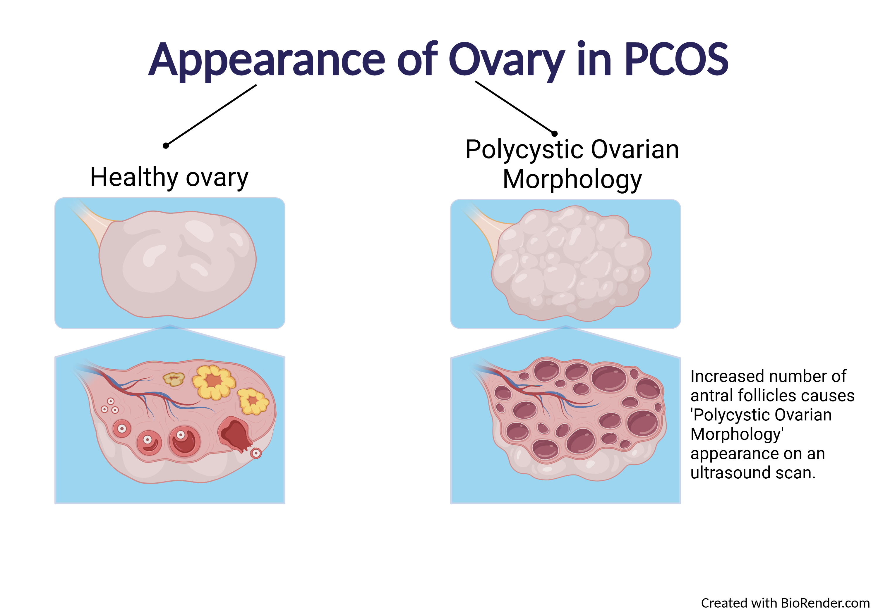
- Increased number of follicles: The ovary is filled with many small, arrested follicles.
- Peripheral Distribution (the "String of Pearls" sign): The follicles are often arranged around the outer edge of the ovary.
- Increased Ovarian Stroma: The central supportive tissue of the ovary appears more prominent and hyperechoic (brighter).
- Enlarged Ovarian Volume: The overall size of the ovary is increased.
The image below provides a helpful schematic, linking the ultrasound appearance to the underlying biology of arrested follicular development that we have discussed in previous lessons.
The Formal Diagnostic Criteria for PCOM
While the visual "string of pearls" is a classic sign, a formal diagnosis of PCOM relies on objective, measurable criteria. These criteria have evolved over time with advances in ultrasound technology.
Let's begin by reviewing the foundational criteria established in 2003.
Ultrasound Assessment in PCOS - Diagnosis
To understand the specifics of the diagnosis, we'll consult a comprehensive review titled 'Ultrasound Assessment in PCOS - Diagnosis'. This paper details the evolution of the diagnostic criteria.
Please read the section titled '3.2. Rotterdam Criteria and Transvaginal Ultrasound'. This section outlines the original 2003 criteria for PCOM, including the specific thresholds for follicle number and ovarian volume. Note the preference for transvaginal ultrasound and the conditions that preclude a correct evaluation.
Based on the 2003 Rotterdam consensus, PCOM is diagnosed if at least one of the following is present in at least one ovary:
- Follicle Number Per Ovary (FNPO): A count of ≥12 follicles measuring 2–9 mm in diameter.
- Ovarian Volume (OV): An ovarian volume of >10 cm³ (or 10 mL).
It's important to note that the presence of a dominant follicle (>10 mm) or a corpus luteum means the ovary cannot be accurately assessed for PCOM in that cycle. The scan is ideally performed in the early follicular phase (days 2-5 of the cycle).
Evolution of the Criteria: Keeping Pace with Technology
Why have these numbers changed over the years? The primary reason is the improvement in ultrasound technology. Modern high-frequency (≥8 MHz) transvaginal probes are much more sensitive and can detect more and smaller follicles than the equipment used in 2003.
Using the old cutoff of ≥12 follicles with new technology would lead to overdiagnosis, as many healthy, ovulatory women would meet this criterion. This led to an update in the international evidence-based guidelines.
Current Guidelines for Diagnosing PCOS - PMC
For the most current recommendations, let's look at the paper 'Current Guidelines for Diagnosing PCOS'. This article provides a clear and concise summary of the updated criteria.
Please read section '5. Ovarian Morphology' and the final 'Current Recommendation' at the end of that section. This text explains the debate around the follicle number threshold and presents the most recent guideline-recommended numbers for diagnosing PCOM with modern ultrasound equipment.
As the resources explain, the 2018 International Guideline updated the PCOM definition for use with modern ultrasound technology:
- Follicle Number Per Ovary (FNPO): A count of ≥20 follicles per ovary.
- Ovarian Volume (OV): The threshold remains >10 cm³.
Important Note: If older ultrasound technology is being used, the original Rotterdam criteria (≥12 follicles) may still be applicable. However, the ≥20 follicle count is the current standard when using high-resolution transvaginal probes.
The following video provides a radiologist's perspective, showing how these criteria are applied in real cases and discussing the evolution from 12 to 20+ follicles.
Ultrasound of Polycystic Ovarian Syndrome
The video 'Ultrasound of Polycystic Ovarian Syndrome' from Radquarters demonstrates the practical application of these criteria with clear case examples.
Please watch from 01:49 to 03:48. Dr. Coble clearly explains the Rotterdam criteria and the subsequent debate and revision of the follicle number cutoff, reinforcing what you've just read.
Interpretation and Important Caveats
Correctly interpreting the ultrasound findings is just as important as obtaining them. There are two critical points to always remember.
1. PCOM is NOT the same as PCOS.
An ultrasound finding of polycystic ovarian morphology is only one part of a three-part definition. A significant percentage of healthy women without any other signs or symptoms have ovaries that meet the ultrasound criteria for PCOM.
To diagnose PCOS, a patient must meet two out of the three Rotterdam criteria:
- Oligo- or anovulation (menstrual dysfunction)
- Clinical and/or biochemical hyperandrogenism
- Polycystic ovarian morphology on ultrasound
2. Ultrasound is not recommended for diagnosis in adolescents.
During the first several years after menarche, it is normal for the ovaries to have a "multicystic" or polycystic appearance as the hypothalamic-pituitary-ovarian (HPO) axis matures. Using ultrasound for diagnosis in this population can lead to significant overdiagnosis. For this reason, international guidelines recommend not using pelvic ultrasound for the diagnosis of PCOS in individuals who are less than 8 years post-menarche.
Test your understanding!
A 28-year-old woman with regular menstrual cycles and no clinical signs of hyperandrogenism (no hirsutism or acne) undergoes a pelvic ultrasound for an unrelated reason. The report notes that her right ovary has a volume of 12 cm³ and contains 24 follicles. Does she have PCOS?
Show answer
No, based on this information, she does not have PCOS. She meets the ultrasound criteria for Polycystic Ovarian Morphology (PCOM). However, she has regular cycles (implying normal ovulation) and no signs of hyperandrogenism. Since she only fulfills one of the three Rotterdam criteria, she cannot be diagnosed with PCOS. This is a classic example of isolated PCOM without the syndrome itself.

Conclusion
Today we have dissected the third diagnostic criterion for PCOS: polycystic ovarian morphology. You now understand what PCOM looks like on an ultrasound and the specific, quantitative criteria used to define it. Most importantly, you can now place this finding in its proper clinical context.
Key Takeaways:
- PCOM is characterized visually by numerous small (2-9mm) follicles, often in a "string of pearls" pattern, with increased central stroma.
- The current formal diagnostic criteria (using modern ultrasound) are a follicle count of ≥20 per ovary OR an ovarian volume of >10 cm³.
- Only one ovary needs to meet the criteria to diagnose PCOM.
- The follicle count threshold was increased from 12 to 20 to account for improvements in ultrasound technology and reduce overdiagnosis.
- PCOM is only one component of the PCOS diagnosis and must be interpreted in the context of the patient's menstrual history and androgen status.
- Ultrasound is not a reliable tool for diagnosing PCOS in adolescents.
Preview of the Next Lesson:
Over the last few lessons, we have systematically covered the clinical presentation, biochemical workup, and ultrasound findings relevant to PCOS. We have also touched on several conditions that can mimic it. In our next lesson, we will synthesize this information to learn how to: Explain the process of differential diagnosis to exclude conditions such as congenital adrenal hyperplasia and androgen-secreting tumors. This will consolidate your diagnostic knowledge and prepare you to understand the broader clinical management of PCOS.