Hello! Welcome to the fourth lesson in our module on the physiology of the normal menstrual cycle.
Introduction
In our last lesson, we examined the dramatic climax of the follicular phase: ovulation. We saw how the LH surge triggers a cascade of molecular events, culminating in the rupture of the Graafian follicle and the release of the oocyte.
But what happens to the follicular structure left behind on the ovary? And what hormonal changes does this set in motion? That brings us to today's learning outcome: to describe the hormonal changes and corpus luteum function during the luteal phase.
We will trace the transformation of the ruptured follicle into a powerful, albeit temporary, endocrine gland called the corpus luteum. We'll explore the hormones it produces and understand how they orchestrate the second half of the menstrual cycle, preparing the body for a potential pregnancy. This phase is defined by the dominance of progesterone, a hormone whose actions are fundamental to reproductive health and central to understanding menstrual cycle abnormalities.
1. From Rupture to Gland: The Formation of the Corpus Luteum
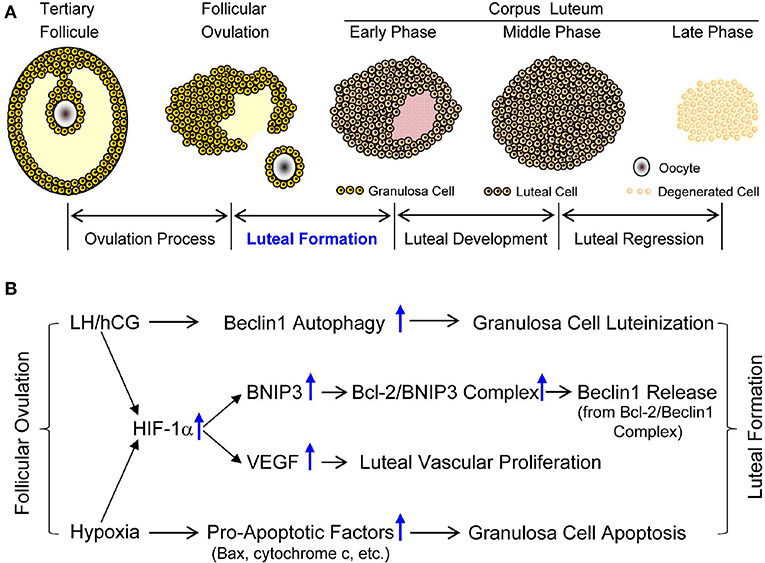
Following the extrusion of the oocyte during ovulation, the remnants of the Graafian follicle—the theca and granulosa cells—begin a rapid and remarkable transformation. Under the influence of LH, these cells undergo luteinization (from the Latin luteus, meaning "yellow").
During this process:
- The cells enlarge and begin to accumulate a yellow pigment called lutein, giving the new structure its name: corpus luteum, or "yellow body."
- The basement membrane that once separated the theca and granulosa cells breaks down.
- Blood vessels from the theca layer invade the granulosa cell layer, creating a rich vascular network. This high degree of vascularization is essential for the corpus luteum to secrete its hormones into the bloodstream effectively.
The resulting structure is a transient endocrine gland, with the former granulosa cells becoming large luteal cells and theca cells becoming small luteal cells, both dedicated to hormone production.
2. The Hormonal Signature of the Luteal Phase
The luteal phase, which lasts for a relatively constant 14 days in most women, is hormonally distinct from the follicular phase. To visualize this shift, let's look at the complete hormonal profile of the menstrual cycle.

The key hormonal products of the corpus luteum are:
- Progesterone: This is the signature hormone of the luteal phase. The luteinized granulosa cells become progesterone-producing powerhouses.
- Estradiol: The corpus luteum continues to produce estrogen, leading to a secondary rise in estradiol levels during the mid-luteal phase.
- Inhibin A: This hormone is specifically produced by the corpus luteum (in contrast to Inhibin B from the follicular phase) and plays a role in regulating FSH.
To understand how the corpus luteum produces these hormones, please watch the following video. It explains the cellular changes and the shift in hormone synthesis after ovulation.
The following clip from Osmosis clearly explains how the luteinized granulosa and theca cells collaborate to produce the characteristic hormones of the luteal phase, with a special focus on the rise of progesterone.
Please watch the video from 07:22 to 08:12. Pay close attention to how the luteinized granulosa cells gain the ability to produce large amounts of progesterone directly from cholesterol.
For a more detailed textual account of this phase, the following reading provides an excellent summary.
The Normal Menstrual Cycle and the Control of Ovulation - NCBI
This chapter from the NCBI bookshelf, 'The Normal Menstrual Cycle and the Control of Ovulation,' provides a concise yet detailed description of the luteal phase.
Please read the section titled 'LUTEAL PHASE'. Focus on understanding the definition and primary function of the corpus luteum, the consistent 14-day duration of this phase, and the pattern of hormone secretion (progesterone and the secondary estradiol rise).
3. The System-Wide Effects of Luteal Hormones
The surge of progesterone, along with estradiol and inhibin A, has profound effects throughout the reproductive system. Its primary mission is to create a welcoming environment for a potential embryo.
a) Negative Feedback on the HPO Axis
Progesterone is a powerful inhibitor of the HPO axis. It acts on the hypothalamus to slow the frequency of GnRH pulses. This, combined with the effects of estradiol and inhibin A on the pituitary, leads to strong suppression of both FSH and LH. This negative feedback is crucial because it prevents the recruitment and development of new follicles during the luteal phase, ensuring that the cycle's focus remains on supporting a potential pregnancy.
b) Preparing the Endometrium: The Secretory Phase
While estrogen in the follicular phase caused the endometrium to grow (proliferate), progesterone now transforms it into a nutrient-rich, receptive bed for implantation. This is called the secretory phase of the uterine cycle. Progesterone's effects include:
- Stimulating Uterine Glands: It causes the glands in the endometrium to mature and secrete a fluid rich in glycogen and other nutrients, sometimes called "uterine milk."
- Increasing Vascularity: It promotes the further development and coiling of the spiral arteries, maximizing blood supply to the uterine lining.
- Thickening Cervical Mucus: It causes the mucus in the cervix to become thick and viscous, forming a plug that blocks sperm from entering the uterus and protects it from infection.
The following video provides a clear explanation of these critical changes to the uterus.
Female Reproductive Cycle | Ovulation & Menstrual Cycle: Overview
This clip from Ninja Nerd effectively illustrates how progesterone acts on the uterus during the luteal phase, transforming it into a secretory lining.
Please watch from 07:55 to 08:38. Focus on the three key changes progesterone induces in the uterus: thickening the lining, developing the spiral arteries, and creating a thick cervical plug.
c) The Thermogenic Effect
Progesterone also acts on the brain's temperature-regulating center in the hypothalamus, causing a slight but sustained increase in basal body temperature (by about 0.3-0.5°C or 0.5-1.0°F). This temperature shift, which persists throughout the luteal phase, is a classic clinical indicator that ovulation has occurred.
Test your understanding!
During the mid-luteal phase (around day 21 of a 28-day cycle), what would you expect the plasma levels of FSH and LH to be, and why?
Show answer
You would expect the levels of both FSH and LH to be very low. This is due to the strong negative feedback exerted by the high levels of progesterone and estradiol produced by the corpus luteum, which suppress GnRH release from the hypothalamus and gonadotropin secretion from the pituitary.
4. The Two Fates of the Corpus Luteum
The corpus luteum is a transient structure with a pre-programmed lifespan. Its ultimate fate depends entirely on one event: whether or not fertilization and implantation occur.
Fate 1: No Pregnancy → Luteolysis
If the oocyte is not fertilized, the corpus luteum has a lifespan of about 14 days. Its survival depends on continuous stimulation from LH. Around 9-11 days after ovulation, it begins to self-destruct in a process called luteolysis (degeneration of the luteum).
- Hormonal Decline: As the corpus luteum degenerates, its production of progesterone, estradiol, and inhibin A plummets.
- Menstruation: This withdrawal of hormonal support, particularly progesterone, causes the spiral arteries in the endometrium to spasm and constrict. The uterine lining becomes ischemic (deprived of blood), breaks down, and is shed, resulting in menstruation.
- A New Beginning: The sharp drop in hormones removes the negative feedback on the HPO axis. GnRH pulses begin to increase in frequency, stimulating the pituitary to release FSH and LH again, which in turn initiates the recruitment of a new cohort of follicles for the next cycle.
- The degenerated corpus luteum becomes an avascular scar tissue called the corpus albicans ("white body"), which is gradually reabsorbed by the ovary over several months.
Fate 2: Pregnancy → hCG Rescue
If fertilization and implantation do occur, the developing embryo begins to produce a hormone called human chorionic gonadotropin (hCG).
- The Rescue Signal: hCG is structurally very similar to LH and binds to the same LH receptors on the corpus luteum. It acts as a powerful rescue signal, overriding the luteolysis program and stimulating the corpus luteum to continue producing progesterone.
- Maintaining Pregnancy: This continued progesterone production (the "progesterone of pregnancy") is absolutely essential for maintaining the uterine lining and sustaining the early embryo.
- Placental Takeover: The hCG-rescued corpus luteum continues this vital function until about 8-12 weeks of gestation, by which time the placenta has developed sufficiently to take over as the primary source of progesterone for the remainder of the pregnancy. After this point, the corpus luteum finally degenerates.
The following resource provides a concise summary of these two distinct pathways.
Anatomy, Ovary Corpus Luteum - NCBI
The article 'Anatomy, Ovary Corpus Luteum' clearly outlines the two possible fates of the corpus luteum.
Please read the section titled 'Structure and Function'. Focus on the two paragraphs that begin with 'The corpus luteum has 2 fates...'. This will reinforce your understanding of the role of hCG and what happens in its absence.
Conclusion
Today we have followed the journey of the post-ovulatory follicle as it transforms into the corpus luteum, a temporary but vital endocrine organ. We have seen how its hormonal output defines the second half of the menstrual cycle.
Key Takeaways:
- After ovulation, the ruptured follicle undergoes luteinization under LH influence to become the corpus luteum.
- The luteal phase is characterized by high levels of progesterone, with a secondary rise in estradiol and the presence of inhibin A, all produced by the corpus luteum.
- These hormones exert strong negative feedback on the HPO axis, suppressing FSH and LH and preventing new follicular development.
- Progesterone transforms the uterine lining into a secretory endometrium, preparing it for implantation.
- If pregnancy does not occur, the corpus luteum degenerates after ~14 days (luteolysis), leading to a drop in hormones that triggers menstruation and restarts the cycle.
- If pregnancy occurs, the embryo produces hCG, which "rescues" the corpus luteum, ensuring continued progesterone production to support the early pregnancy.
Preview of the Next Lesson:
In this lesson and the first lesson of this module, we have discussed the uterine changes that happen in response to ovarian hormones—the proliferative phase and the secretory phase. In our next lesson, we will synthesize this information to fulfill the learning outcome: "Correlate the endometrial changes (proliferative and secretory phases) with the ovarian cycle." We will place these two uterine phases side-by-side to fully appreciate how they mirror the follicular and luteal phases of the ovarian cycle.