Hello! In our last lesson, we explored the general blueprint for all synovial joints, focusing on the articular cartilage, joint capsule, and synovial fluid that enable free movement. Now, we'll build on that foundation to see how this basic design is adapted into different forms to serve various functions throughout the body.
This lesson directly addresses your goal of classifying the six types of synovial joints. We'll categorize them based on the shape of their articulating surfaces—a feature that directly determines the kind of movement each joint can perform. Understanding this link between structure and function is essential for your A&P exam and for clinical practice, where you'll assess patient mobility and joint-related conditions.
From Shape to Movement: Classifying Synovial Joints
While all synovial joints share a common structure, the specific shape of the bones' articulating surfaces varies greatly. This shape is the primary factor that dictates the joint's range and type of motion. We can group these joints into three functional categories based on their axes of movement:
- Uniaxial: Movement in one plane (e.g., like a door hinge).
- Biaxial: Movement in two planes.
- Multiaxial: Movement in all three planes.
To begin, let's watch a video that introduces the six joint types and provides a convenient mnemonic to help you remember them for your exam. The video organizes the joints using the three axial categories, which we will use as the framework for this lesson.
This segment from "Synovial Joints" by Dr. Matt & Dr. Mike will introduce the six types of joints and a helpful way to remember them.
Watch from the beginning until the introduction of the axes. Pay close attention to the mnemonic: Prince Harry Pooled Charles's Saddle Bag. This will help you recall the six types: Planar, Hinge, Pivot, Condylar, Saddle, and Ball-and-socket.
Now, let's examine each of these types in more detail, following the uniaxial, biaxial, and multiaxial structure.
1. Uniaxial Joints (Movement in One Plane)
These joints allow motion around a single axis.
Hinge Joint
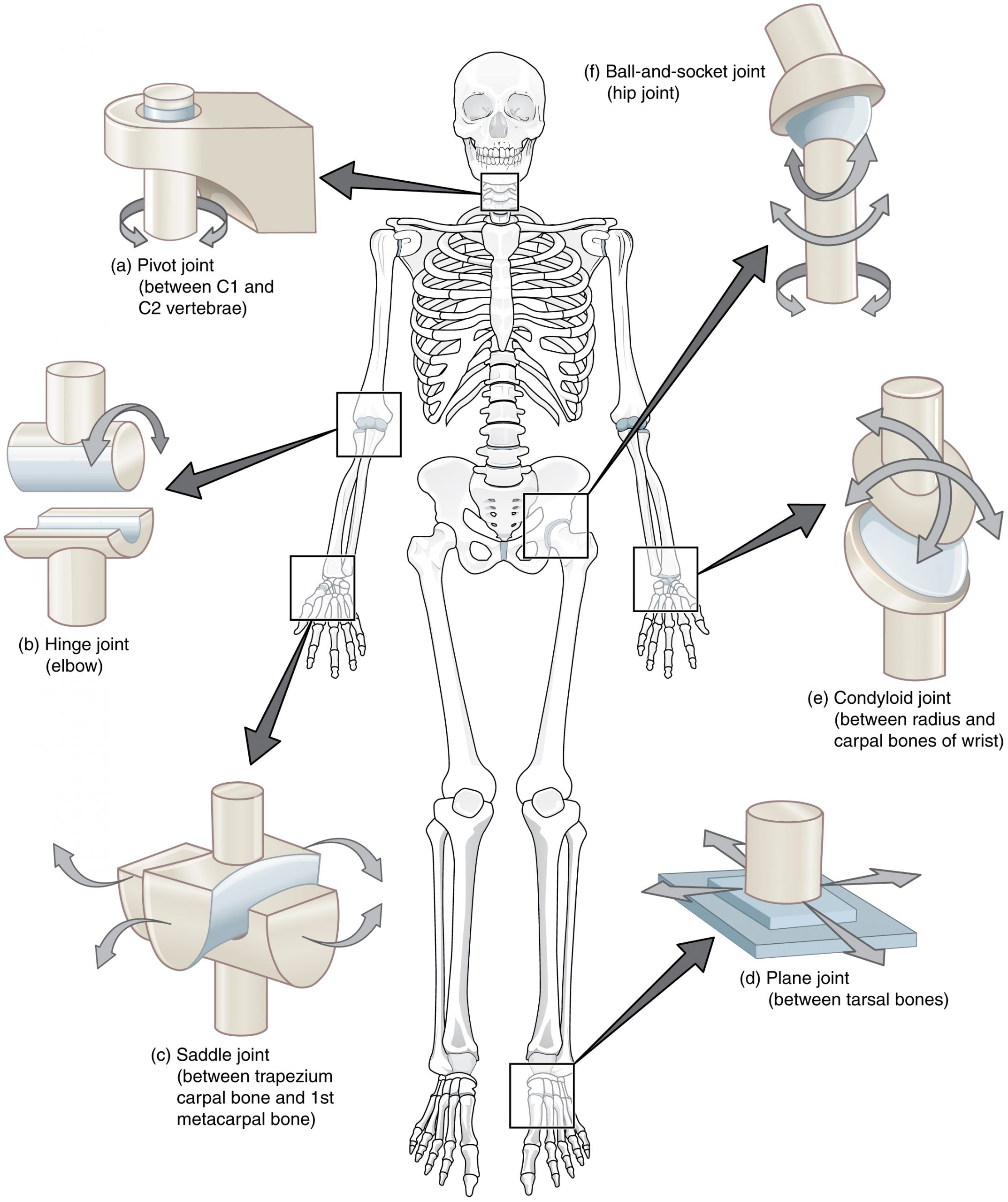
Just as its name suggests, a hinge joint works like the hinge on a door, allowing movement back and forth in one direction. In a hinge joint, the convex surface of one bone fits into the concave surface of another.
- Movement: Allows only for flexion and extension.
- Examples: The elbow joint (articulation between the humerus and ulna), the knee joint, and the interphalangeal joints of your fingers and toes.
Pivot Joint
In a pivot joint, a rounded portion of one bone rotates within a ring formed by another bone and a ligament. This design permits rotation around a central axis.
- Movement: Allows only for rotation.
- Examples:
- The atlantoaxial joint between the first (atlas) and second (axis) cervical vertebrae. This joint allows you to shake your head "no."
- The proximal radioulnar joint, where the head of the radius pivots within a ring formed by the ulna. This joint allows you to turn your palm up (supination) and down (pronation).
To see how the structure of these joints creates their specific movements, the following video offers excellent visualizations.
The 6 Types of Joints - Human Anatomy for Artists
The video "The 6 Types of Joints" by Proko, while intended for artists, provides exceptionally clear animations of how these joints function.
Watch the section on Hinge joints, followed by the section on Pivot joints. Notice how the shape of the bones restricts movement to a single plane or axis.
2. Biaxial Joints (Movement in Two Planes)
These joints allow motion around two axes, providing more freedom than uniaxial joints.
Condylar (or Ellipsoid) Joint
Here, an oval-shaped convex end of one bone articulates with an elliptical cavity of another.
- Movement: Allows flexion/extension and abduction/adduction (side-to-side movement).
- Examples: The radiocarpal (wrist) joint and the metacarpophalangeal joints (the knuckles at the base of your fingers). These joints allow you to wave your hand and spread your fingers apart.
Saddle Joint
Saddle joints feature two bones that both have concave and convex surfaces, fitting together like a rider on a saddle. This unique shape allows for a greater range of motion than a condylar joint.
- Movement: Allows flexion/extension, abduction/adduction, and circumduction.
- Example: The first carpometacarpal joint at the base of the thumb. This is the joint that gives us our opposable thumb, a crucial element of human dexterity.
3. Multiaxial Joints (Movement in Multiple Planes)
These joints offer the greatest degree of motion.
Plane (or Gliding) Joint
Plane joints are formed by the articulation of two flat or slightly curved bone surfaces. While they can technically move in multiple directions, the movement is typically small and limited by surrounding ligaments.
- Movement: Allows for gliding or sliding motions.
- Examples: The intercarpal joints in the wrist, the intertarsal joints in the ankle, and the joints between vertebrae.
Ball-and-Socket Joint
This joint has the most freedom of all synovial joints. A spherical "ball" (the head of one bone) fits into a cup-like "socket" of another.
- Movement: Allows for flexion/extension, abduction/adduction, and rotation.
- Examples: The shoulder (glenohumeral) joint and the hip joint. You may notice in your work at the retirement home that these joints are common sites of injury (shoulder dislocation, hip fracture). This is due to a fundamental trade-off: the shoulder's shallow socket provides great mobility but less stability, while the hip's deep socket provides stability for weight-bearing but limits mobility.
The image below provides a fantastic summary of all six joint types and their locations in the body.
To solidify your understanding, please read the following sections from the "Anatomy & Physiology" textbook. They provide concise, formal definitions and examples for each joint type we have discussed.
Synovial Joints – Anatomy & Physiology
This resource offers clear, systematic descriptions of each joint type, reinforcing what we've covered.
Please read the following six subsections under the main heading "Types of Synovial Joints": Pivot Joint Hinge Joint Condyloid Joint Saddle Joint Plane Joint Ball-and-Socket Joint Focus on connecting the description of the bone shapes to the functional classification (uniaxial, biaxial, or multiaxial).
Conclusion
In this lesson, we've systematically classified the six types of synovial joints based on the shape of their articulating surfaces. This structural classification is key to understanding the function of our skeleton, as shape directly determines the movements a joint can perform.
Key Takeaways:
- Synovial joints are classified by shape into six types: plane, hinge, pivot, condylar, saddle, and ball-and-socket.
- This classification can be organized by axes of movement:
- Uniaxial: Hinge (elbow) and Pivot (atlantoaxial joint).
- Biaxial: Condylar (wrist) and Saddle (thumb).
- Multiaxial: Plane (intercarpal bones) and Ball-and-Socket (hip, shoulder).
- Remembering one clear example for each type is crucial for your exam.
Having learned to identify the different types of joints, our next step is to learn the precise anatomical language used to describe the movements they produce. In the next lesson, we will define terms like flexion, extension, abduction, and adduction.