Welcome back! In our last lesson, we saw how a signal from a motor neuron creates an electrical impulse, an action potential, on the surface of the muscle fiber. But how does this surface-level event trigger a contraction deep within the muscle? Today, we will bridge that gap.
Your learning goal is to explain the process of excitation-contraction coupling, linking the action potential to the release of calcium ions. We'll follow the action potential as it dives into the muscle cell and activates a sophisticated mechanism to release a flood of calcium, the critical chemical messenger that initiates the entire contractile process. This is the "coupling" that turns an electrical "excitation" into the chemical signal for "contraction."
From the Surface to the Interior
A muscle fiber is a large cell packed with myofibrils. For a contraction to be coordinated, the signal—the action potential—must penetrate deep into the cell's interior almost instantly. This is achieved by a specialized network of membranes.
- Transverse Tubules (T-tubules): These are deep, tube-like invaginations of the sarcolemma (the muscle cell membrane) that extend into the sarcoplasm. Think of them as tunnels that carry the electrical signal from the surface to the inside of the cell.
- Sarcoplasmic Reticulum (SR): This is an intricate, web-like membrane system, similar to the endoplasmic reticulum of other cells. Its primary job is to store a high concentration of calcium ions ().
- The Triad: Where a T-tubule runs between two enlarged portions of the SR, called terminal cisternae, a structure known as a triad is formed. This three-part junction is the anatomical basis of excitation-contraction coupling.
The following short video provides an excellent visual review of these structures and sets the stage for the process we are about to explore.
Excitation Contraction Coupling | Skeletal Muscle Contraction | Cross Bridge Cycling | Myology
This video from Byte Size Med, "Excitation Contraction Coupling," will help you visualize the key anatomical players.
Watch the first two minutes, from the beginning to this overview. Focus on how the T-tubules and the sarcoplasmic reticulum are arranged relative to the myofibrils.
The Molecular Machinery of Calcium Release
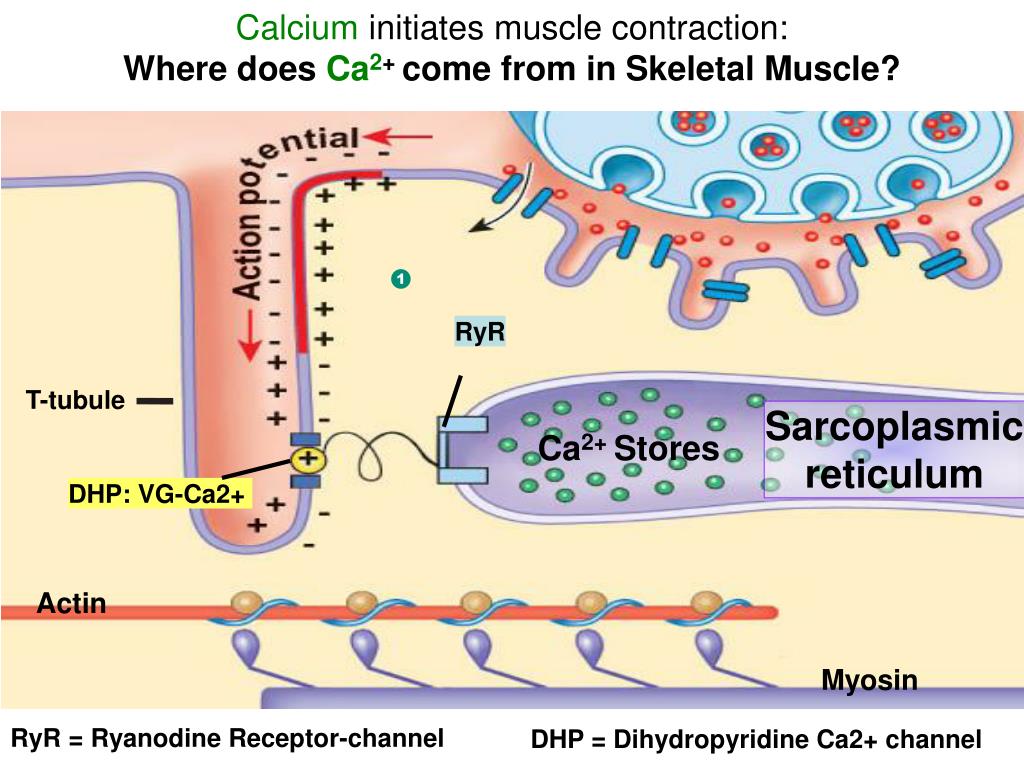
The critical events of excitation-contraction coupling occur at the triad. Embedded within the membranes of the T-tubule and the sarcoplasmic reticulum are two key proteins that work together as a molecular switch.
- Dihydropyridine Receptor (DHPR): Located on the membrane of the T-tubule, this protein is a voltage-sensitive channel. When the action potential travels down the T-tubule, the change in membrane voltage causes the DHPR to change its shape. It acts as a voltage sensor.
- Ryanodine Receptor (RyR): Located on the membrane of the sarcoplasmic reticulum, this protein is a calcium channel. In skeletal muscle, it is physically linked to the DHPR.
The image below provides a close-up view of this crucial interface.
To understand the specifics of this interaction, please read the following section from the "The Cross-Bridge Cycle and Excitation-Contraction Coupling" article.
The Cross-Bridge Cycle and Excitation-Contraction Coupling
This text describes the interaction between the DHP receptor and the Ryanodine receptor.
Please read the section titled Calcium Release. Pay close attention to how a change in the DHP receptor directly causes the Ryanodine receptor to open.
The Coupling Event: From Excitation to Calcium Release
Now, let's put the pieces together to see the full sequence of events.
- An action potential, generated at the neuromuscular junction, propagates across the sarcolemma and down the T-tubules.
- The arrival of the action potential at the triad causes the voltage-sensitive DHP receptors to change their conformation.
- This conformational change in the DHPRs, due to their physical linkage, pulls open the "gate" of the RyR channels on the sarcoplasmic reticulum.
- Calcium ions (), which are stored at very high concentrations inside the SR, rush out through the open RyR channels and into the sarcoplasm, following their steep concentration gradient.
This rapid and massive increase in sarcoplasmic concentration is the central event of excitation-contraction coupling. This is the "on" switch for muscle contraction.
The following video segment provides a clear animation of this exact process.
Excitation Contraction Coupling | Skeletal Muscle Contraction | Cross Bridge Cycling | Myology
The "Excitation Contraction Coupling" video will now show you the coupling process in action.
Watch the segment from this section on calcium release. Notice how the arrival of the action potential leads directly to the opening of the SR channels and the flood of calcium into the sarcoplasm.
Resetting the System: Muscle Relaxation
For a muscle to relax, the sarcoplasmic calcium concentration must be brought back down to its low resting level. If calcium remained high, the muscle would stay contracted. This "off" switch is just as important as the "on" switch.
This is accomplished by a powerful active transport system:
- SERCA Pumps (Sarcoplasmic/Endoplasmic Reticulum -ATPase): These protein pumps are embedded in the membrane of the SR. They continuously use ATP to pump from the sarcoplasm back into the SR, against its concentration gradient.
- Once signaling from the motor neuron stops, the action potentials cease. The RyR channels close, and the SERCA pumps quickly sequester the , causing the muscle fiber to relax.
Inside the SR, a protein called calsequestrin binds to the calcium ions, which helps to lower the free concentration within the SR. This reduces the concentration gradient that the SERCA pumps must work against, making the process of calcium reuptake more efficient.
The final segment of the video explains this relaxation phase.
Excitation Contraction Coupling | Skeletal Muscle Contraction | Cross Bridge Cycling | Myology
This final clip explains how the muscle resets after a contraction.
Watch from the section on relaxation to understand the role of the SERCA pump and the importance of removing calcium from the sarcoplasm.
The entire process of excitation-contraction coupling is summarized beautifully in the diagram below.

Conclusion
In this lesson, we have connected the electrical signal on the muscle cell membrane to the chemical trigger for contraction. You can now explain the crucial process of excitation-contraction coupling.
Key Takeaways:
- Excitation-Contraction (EC) coupling is the process that links the action potential (excitation) to the release of calcium ions from the sarcoplasmic reticulum.
- T-tubules are extensions of the sarcolemma that carry the action potential into the muscle fiber's interior.
- At the triad, a voltage-sensitive DHP receptor on the T-tubule is mechanically linked to a Ryanodine receptor (a channel) on the SR.
- The action potential causes the DHP receptor to change shape, which pulls open the RyR channel, releasing a flood of into the sarcoplasm.
- For relaxation, SERCA pumps use ATP to actively transport back into the SR.
We've now unleashed the key that will unlock the contractile machinery. In our next lesson, we will examine exactly what this flood of calcium does, exploring the roles of troponin and tropomyosin in the cross-bridge cycle and the sliding filament mechanism of muscle contraction.