Welcome back. In our previous lesson, we examined the static anatomy of the sarcomere, the fundamental contractile unit of muscle. We now have a blueprint of the "machinery" responsible for contraction. Today, we'll focus on how this machinery receives its command to act.
Your learning goal is to describe the sequence of events at the neuromuscular junction that initiates a muscle action potential. This is a crucial step in physiology, as it's the bridge between the nervous system and the muscular system. We will trace the path of a nerve signal as it jumps the gap to the muscle fiber, transforming from an electrical signal to a chemical one, and back to an electrical signal again. This process is the essential "on switch" for muscle contraction.
The Anatomy of the Neuromuscular Junction
Before we dive into the sequence of events, let's first familiarize ourselves with the key structures involved. The neuromuscular junction (NMJ) is a specialized synapse—a point of communication—between a motor neuron and a skeletal muscle fiber.
The image below provides a detailed map of the NMJ. We will refer back to the numbered steps in this diagram as we walk through the process.
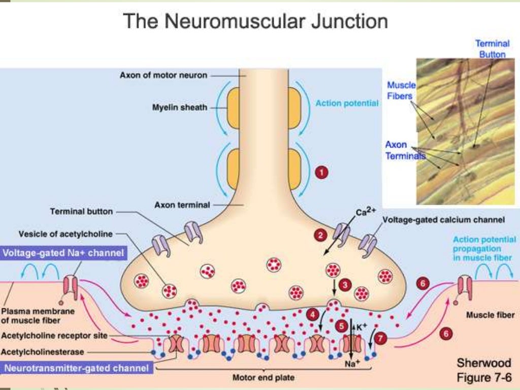
The NMJ consists of three primary parts:
- The Presynaptic Terminal: The enlarged end of the motor neuron's axon. It contains synaptic vesicles filled with the neurotransmitter acetylcholine (ACh). Its membrane is embedded with voltage-gated calcium channels.
- The Synaptic Cleft: The tiny, fluid-filled space separating the neuron from the muscle fiber. The electrical signal cannot cross this gap directly.
- The Motor End Plate: A specialized region of the muscle fiber's membrane (the sarcolemma). It is rich in acetylcholine receptors and features deep folds to increase surface area.
To solidify your understanding of these structures, please read the following brief sections from the Kenhub article "Neuromuscular junction: Structure and function."
Neuromuscular junction: Structure and function
This article provides a clear textual description of the components of the NMJ and their roles.
Focus on these key areas: First, read the section under the heading "Components" to get an overview of the three main parts. Next, review the two short sections titled "Synaptic end bulb" and "Motor end plate" for details on the pre- and post-synaptic structures. Finally, read the section under "Synaptic cleft" to understand the critical role of the enzyme it contains.
The Sequence of Events: From Nerve Impulse to Muscle Action Potential
Now that we have set the stage, let's explore the sequence of events that unfolds when a nerve impulse arrives at the NMJ. This process converts the electrical signal of the neuron into an electrical signal in the muscle fiber.
The following video from Byte Size Med provides a fantastic, step-by-step walkthrough of this entire process.
Neuromuscular Junction | NMJ | Neuromuscular transmission | Myology | Nerve Muscle Physiology
This video, "Neuromuscular Junction," clearly explains the sequence of neurotransmission.
Please watch the following segments carefully: Step 1: Signal Arrival and ACh Release. Watch from this first section, which shows how the arrival of an action potential at the axon terminal triggers the release of acetylcholine. Step 2: Generating the Muscle Signal. Continue with the next part, from this segment on the end plate potential. This is the core of the process, where acetylcholine binding leads to the generation of a new action potential on the muscle fiber. Step 3: Terminating the Signal. Finally, watch the brief section from this part about signal cleanup, which explains how the signal is switched off.
Let's summarize these crucial steps, referencing the numbered events in the diagram shown earlier:
-
Action Potential Arrives: An electrical impulse travels down the axon of the motor neuron to the presynaptic terminal. (See #1 in the diagram).
-
Calcium Influx: The action potential causes voltage-gated calcium () channels on the presynaptic terminal to open. Since the concentration of is much higher outside the cell, calcium ions rush into the terminal. (See #2).
-
ACh Release: The influx of acts as a signal, causing the synaptic vesicles to fuse with the presynaptic membrane and release acetylcholine (ACh) into the synaptic cleft via exocytosis. (See #3).
-
ACh Binds to Receptors: ACh molecules diffuse across the synaptic cleft and bind to ACh receptors on the motor end plate of the muscle fiber. (See #4).
-
Opening of Ligand-Gated Channels: These ACh receptors are ligand-gated cation channels. The binding of two ACh molecules causes the channel to open.
-
End-Plate Potential (EPP): The open channels allow sodium ions () to rush into the muscle fiber, and a smaller amount of potassium ions () to exit. The large influx of positive sodium ions causes the membrane potential at the motor end plate to become less negative, a process called depolarization. This localized voltage change is the end-plate potential. (See #5).
-
Generation of a Muscle Action Potential: The EPP is a graded potential. If it is strong enough to reach a critical threshold value, it triggers the opening of nearby voltage-gated sodium channels on the sarcolemma. The massive influx of through these channels generates a self-propagating electrical impulse—the muscle action potential—that spreads across the entire surface of the muscle fiber. (See #6).
-
Termination of the Signal: To prevent constant contraction, the signal must be terminated quickly. This is accomplished by the enzyme acetylcholinesterase (AChE), which resides in the synaptic cleft. AChE rapidly breaks down ACh into acetyl and choline, which cannot activate the receptors. The choline is then transported back into the axon terminal to be recycled for future ACh synthesis. (See #7 in the diagram, showing ACh breakdown and ion movement ceasing).
As a nursing student, it's useful to know that disruptions in this finely tuned process can lead to significant clinical conditions. For instance, in Myasthenia Gravis, the body's immune system mistakenly attacks and destroys acetylcholine receptors on the motor end plate. This impairs the transmission of the signal, leading to muscle weakness.
Conclusion
In this lesson, we have detailed the critical steps that translate a command from the nervous system into an excitatory signal on a muscle fiber. You can now describe the entire sequence at the neuromuscular junction, from the arrival of a nerve impulse to the generation of a muscle action potential.
Key Takeaways:
- The NMJ is a synapse composed of the presynaptic terminal, synaptic cleft, and motor end plate.
- An electrical signal (action potential) in a neuron triggers the release of a chemical signal (acetylcholine) into the synaptic cleft.
- This chemical signal binds to receptors on the muscle fiber, causing an electrical signal (end-plate potential) at the motor end plate.
- If the end-plate potential reaches threshold, it initiates a full muscle action potential that propagates along the sarcolemma.
- The signal is terminated by the enzyme acetylcholinesterase, ensuring precise control over muscle contraction.
We have successfully generated the "go" signal on the muscle fiber's surface. In our next lesson, we will follow this action potential as it dives deep into the muscle fiber to trigger the release of calcium, a process known as excitation-contraction coupling, which directly leads to the sliding of filaments within the sarcomere.