In our last lesson, we built a foundational map of the skull by identifying its 22 individual bones, separating them into the cranium and the facial skeleton. Now, we'll focus on how the large, flat bones of the cranium are held together. This lesson examines the immovable joints of the skull, which are crucial for protecting the brain.
Your learning outcome for this session is to identify the major sutures of the skull: the sagittal, coronal, squamous, and lambdoid sutures. By the end of this lesson, you will be able to locate each of these sutures and name the specific cranial bones they unite, adding another layer of detail to your anatomical understanding of the axial skeleton.
What is a Suture?
Before we identify the specific sutures, it's important to understand what they are and why they are structured in such a unique way. The bones of the cranium aren't simply fused together; they are connected by a specific type of joint.
7.2 The Skull – Anatomy & Physiology 2e
This reading from the Anatomy & Physiology 2e textbook provides a concise definition of a suture and its functional importance.
Please read the two short paragraphs under the heading "Sutures of the Skull," starting with the definition of a suture. Pay attention to the type of connective tissue involved and the reason for the irregular, interlocking lines.
As you just read, a suture is an immobile joint filled with dense, fibrous connective tissue. Their complex, twisting paths are not random; they serve to tightly interlock the cranial bones, much like the pieces of a jigsaw puzzle, which significantly increases the strength of the skull.

Identifying the Four Major Sutures
There are many sutures in the skull, but for your exam and foundational knowledge, we will focus on the four largest and most prominent ones. The following diagram provides an excellent overview from both a superior (top) and lateral (side) perspective.
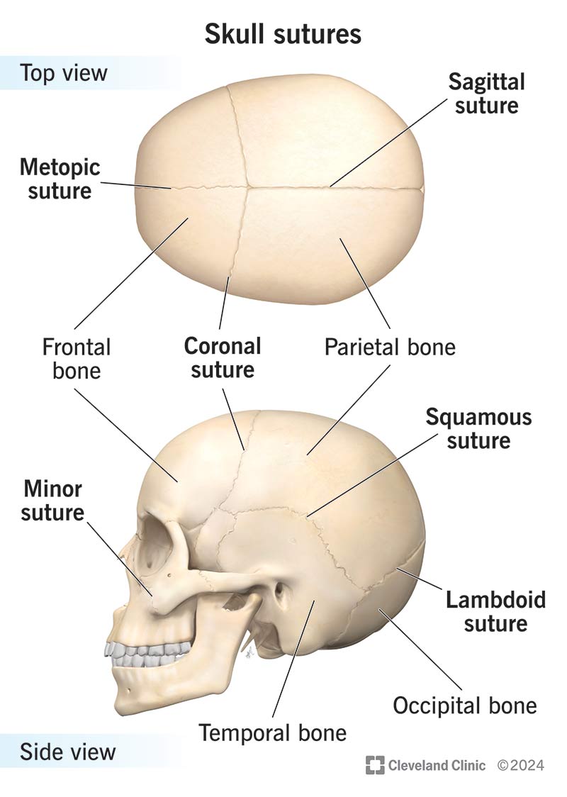
Now, let's take a guided tour to see these sutures on a skull model. The following video will help you visualize their location and orientation from multiple angles.
Skull bones, sutures and landmarks
This video from "The Noted Anatomist" provides an excellent tour of the skull, focusing specifically on the sutures from multiple perspectives. The narrator also explains the origin of the names, which can be helpful memory aids.
Please watch the following segments: Lateral view: This covers the coronal, squamous, and lambdoid sutures. Posterior view: This focuses on the lambdoid and sagittal sutures, explaining the "lambda" shape. Superior view: This provides a final review of the coronal, sagittal, and lambdoid sutures from the top down.
A Detailed Breakdown
Having seen the sutures visually, let's solidify your knowledge with detailed descriptions. It is crucial to be able to name not just the suture, but also the bones it connects.
7.2 The Skull – Anatomy & Physiology 2e
Now let's reinforce what you saw in the video with detailed text descriptions from Anatomy & Physiology 2e. This will help you commit the names and locations to memory for your exam.
Please read the paragraph that describes the four major sutures, starting with the coronal and sagittal sutures and continuing through the descriptions of the lambdoid and squamous sutures. As you read, mentally trace each suture on the "Skull sutures" diagram we looked at earlier.
To help you organize this information for efficient studying, here is a summary table. This format is excellent for quick review.
| Suture | Location & Orientation | Bones United |
|---|---|---|
| Coronal Suture | Runs from side to side across the skull, separating the front from the back. It lies in the coronal (or frontal) plane. | Frontal bone with the left and right Parietal bones. |
| Sagittal Suture | Runs front-to-back along the midline on top of the skull. It lies in the sagittal plane. | The left and right Parietal bones. |
| Lambdoid Suture | Forms an inverted "V" shape on the posterior aspect of the skull. Its name comes from the Greek letter lambda (Λ). | Occipital bone with the left and right Parietal bones. |
| Squamous Suture | Arches along the lateral side of the skull, superior to the ear. It is named for the flat, "scaly" squamous portion of the temporal bone. | Temporal bone with the Parietal bone on each side. |
Check Your Understanding
To prepare for your exam, it's a great practice to test your recall.
7.2 The Skull – Anatomy & Physiology 2e
Let's use a review question from the textbook to check your understanding.
Find the question that asks you to identify the major sutures. First, try to answer it yourself without looking at your notes. Then, click the "Reveal" button provided in the resource to check your answer and reinforce the material.
Conclusion
In this lesson, you have learned how the protective case for the brain, the cranium, is assembled. We moved from knowing the individual bones to understanding how they are joined by strong, immovable fibrous joints called sutures.
Key Takeaways:
- A suture is an immobile fibrous joint that tightly interlocks the bones of the skull.
- The Coronal suture unites the frontal bone with the two parietal bones.
- The Sagittal suture unites the right and left parietal bones at the midline.
- The Lambdoid suture unites the occipital bone with the two parietal bones.
- The Squamous suture unites the temporal bone with the parietal bone on each side of the skull.
You have now mapped out the major bones of the cranium and the sutures that bind them. In the next lesson, we will continue our exploration of the skull by identifying its key openings, or foramina. These passageways are critical, as they allow major nerves and blood vessels to enter and exit the cranial cavity, connecting the brain to the rest of the body.