Welcome to the first lesson in our new module on the Axial Skeleton. In the previous module, we explored the microscopic world of bone tissue, understanding how bones grow, remodel, and manage the body's calcium balance. Now, we shift our focus from the cellular level to the gross anatomy of the skeleton itself, beginning with the structure that houses our most vital organ: the skull.
This lesson addresses the first step in understanding the axial skeleton: identifying the major bones of the cranium and face. The skull is more than just a single bone; it's an intricate puzzle of 22 bones that fit together to protect the brain and form the structure of our face. By the end of this lesson, you will be able to name and locate these bones, creating a foundational map for our subsequent study of the skull's more detailed features.
An Overview of the Skull
The skull is the body's most complex bony structure. It is traditionally divided into two main parts: the cranium (or brain case), which encloses the brain, and the facial skeleton, which forms the framework of the face.
The Skull | Anatomy and Physiology I
This introductory section from Lumen Learning's "Anatomy and Physiology I" textbook provides the formal definitions and categories for the bones of the skull.
Please read from the beginning of the section, starting with the definition of the cranium. This will establish the fundamental division between the cranial and facial bones.
As you've just read, the skull consists of 22 bones. Eight of these form the cranium, and fourteen form the face. The only movable bone is the mandible, or lower jaw.
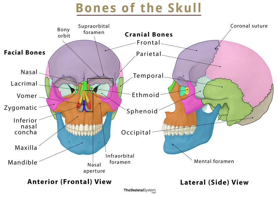
Let's use a diagram as our roadmap for this lesson. The image below shows the skull from the front (anterior) and side (lateral) views, with the different bones color-coded for clarity. Refer back to this image as we discuss each bone.
The Cranial Bones: Protecting the Brain
The eight cranial bones form the protective vault and base of the skull that enclose the brain. They include single bones and paired bones.
- Single bones: Frontal, Occipital, Sphenoid, Ethmoid
- Paired bones: Parietal (left and right), Temporal (left and right)
The following video provides an excellent visual walkthrough, identifying each of these bones on a 3D model from both outside and inside the skull.
This video, "Bones of the Cranium and Face" from the Anatomy Hero channel, clearly demonstrates the location of each cranial bone.
First, watch the segment showing the external view of the cranial bones. Then, continue with the internal view, which gives a unique perspective on the sphenoid, ethmoid, and occipital bones.
Now that you have a visual understanding of where these bones are located, let's solidify that knowledge with a more detailed description of each one.
The Skull | Anatomy and Physiology I
This section of the Lumen Learning text provides detailed descriptions of the eight cranial bones.
Please read the section titled Bones of the Brain Case. Focus on identifying the key features and location of each of the eight bones: Parietal Bone Temporal Bone Frontal Bone Occipital Bone Sphenoid Bone Ethmoid Bone As you read, try to locate each bone and its main described features on the "Bones of the Skull" diagram we looked at earlier.
The sphenoid and ethmoid bones can be tricky because they are mostly hidden internally. The sphenoid is often called the "keystone" because it articulates with all other cranial bones. The ethmoid is a delicate bone found between the eyes that forms part of the nasal cavity and the orbits.
The Facial Bones: Forming the Face
The fourteen facial bones provide the framework for our facial structures, support the teeth, and form the nasal cavity and eye orbits. Like the cranium, this group contains both single and paired bones.
- Single bones: Mandible, Vomer
- Paired bones: Maxilla, Zygomatic, Nasal, Lacrimal, Palatine, Inferior nasal concha
Let's return to the "Bones of the Cranium and Face" video to see where these bones are located.
The narrator will now walk you through the facial bones using the 3D model.
Watch the portion of the video from the facial bones segment. The narrator does an excellent job of explaining the position of each bone relative to its neighbors.
With a visual map of the facial skeleton, you're ready to dive into the details. The following reading describes the location and primary features of each of the 14 facial bones.
The Skull | Anatomy and Physiology I
This final reading covers the facial skeleton in detail.
Please read the section titled Facial Bones of the Skull. Systematically go through each of the following bones: Maxillary Bone (Maxilla) Palatine Bone Zygomatic Bone (Cheekbone) Nasal Bone Lacrimal Bone Inferior Nasal Conchae Vomer Bone Mandible (Lower Jaw)
A Tool for Memorization
Given your upcoming exam, memorizing these 22 bones can be challenging. Mnemonics are a great tool for quick recall. The following video offers some popular mnemonics for both the cranial and facial bones.
Skull Bones Mnemonic (Cranial and Facial Bones) | Anatomy and Physiology
The video "Skull Bones Mnemonic" by RegisteredNurseRN is designed specifically for memorization.
Watch from the beginning to the end. The mnemonics "Ethan's Fried Spanish Pasta Tempted Octavia" for the cranial bones and "My Mouth's Palate Never Liked Zucchini In Vinegar" for the facial bones may be helpful study aids.
Conclusion
In this lesson, we have moved from the general structure of osseous tissue to the specific anatomy of the skull. You have learned to identify the 22 bones that compose it, dividing them into their functional groups: the 8 bones of the cranium and the 14 bones of the face.
Key Takeaways:
- The skull is composed of two main units: the cranium (brain case) and the facial skeleton.
- The cranium consists of 8 bones:
- Single: Frontal, Occipital, Sphenoid, Ethmoid
- Paired: Parietal, Temporal
- The facial skeleton consists of 14 bones:
- Single: Mandible, Vomer
- Paired: Maxilla, Zygomatic, Nasal, Lacrimal, Palatine, Inferior Nasal Conchae
You now have a solid anatomical map of the skull's components. In our next lesson, we will build upon this foundation by examining how these bones connect. We will identify the major sutures of the skull, which are the immovable joints that hold the cranial bones together, providing the cranium with its strength and rigidity.