Welcome back. In our last lesson, we focused on the distinct features of the vertebrae in each spinal region. We paid special attention to the thoracic vertebrae and their unique costal facets, the attachment points for the ribs. Today, we will complete our tour of the axial skeleton by building upon that knowledge to examine the entire thoracic cage.
Your goal for this lesson is to identify the components of the thoracic cage, including the sternum and all types of ribs (true, false, and floating). For a future nurse, this anatomy is not just a list of names to memorize; it forms the basis for critical clinical skills. Understanding the precise layout of the ribs and sternum is essential for accurately placing a stethoscope to listen to heart and lung sounds, performing CPR correctly, and assessing chest injuries.
The Sternum: The Central Anchor
The thoracic cage forms the bony framework of your chest, protecting vital organs like the heart and lungs. At the very front and center of this cage lies the sternum, or breastbone. It serves as the anterior anchor for the ribs. The sternum itself is not one solid bone but is composed of three distinct parts.
To get a clear overview of the sternum and its clinically important landmarks, let's watch a brief video.
Thoracic Anatomy: Complete Guide to Skeleton, Sternum & Ribs | Lecturio Medical
This video from Lecturio Medical provides a concise introduction to the anatomy of the sternum. The presenter clearly points out the main parts and landmarks that you will need to identify.
Please watch the first part of the video, from the beginning up to the discussion on ribs. As you watch, focus on identifying these three key structures and two important landmarks: Manubrium (the superior part) Body (the middle and largest part) Xiphoid process (the inferior tip) Jugular (suprasternal) notch (the palpable dip at the top) Sternal angle (the ridge between the manubrium and body)
As the video explained, these parts are not just abstract anatomical terms. They have direct clinical applications:
- The sternal angle (also called the Angle of Louis) is a crucial landmark. It's a palpable ridge on the front of the chest that marks the point where the second rib attaches. Since the first rib is difficult to feel, clinicians use the sternal angle as the starting point to count ribs, which is essential for locating specific intercostal spaces for auscultation or other procedures.
- The xiphoid process, the small cartilaginous tip at the bottom of the sternum, is another vital landmark, particularly during cardiopulmonary resuscitation (CPR). Correct hand placement is crucial to avoid fracturing the xiphoid process, which could potentially damage the liver.
- The manubrium also features the clavicular notches, where the clavicles (collarbones) attach. This joint, the sternoclavicular joint, is our first link to the appendicular skeleton, which we will begin studying in the next module.
The Ribs: A Protective Cage
Now let's turn to the ribs themselves. Humans have 12 pairs of ribs, all of which articulate posteriorly with the 12 thoracic vertebrae we studied previously. However, their connection at the front of the body varies, and this variation is the basis for their classification.
The bony part of each rib does not extend all the way to the sternum. Instead, most ribs are connected to the sternum by a flexible bridge of costal cartilage. The way this cartilage attaches determines whether a rib is classified as "true," "false," or "floating."
The following reading provides a clear, structured explanation of these classifications.
The Thoracic Cage | Anatomy and Physiology I
This section from a Lumen Learning course on Anatomy and Physiology breaks down the classification of ribs based on their attachment to the sternum.
Please read the section titled Rib Classifications. Focus on understanding the precise definition for each of the three groups.
To summarize and reinforce what you've just read, let's break down the categories:
-
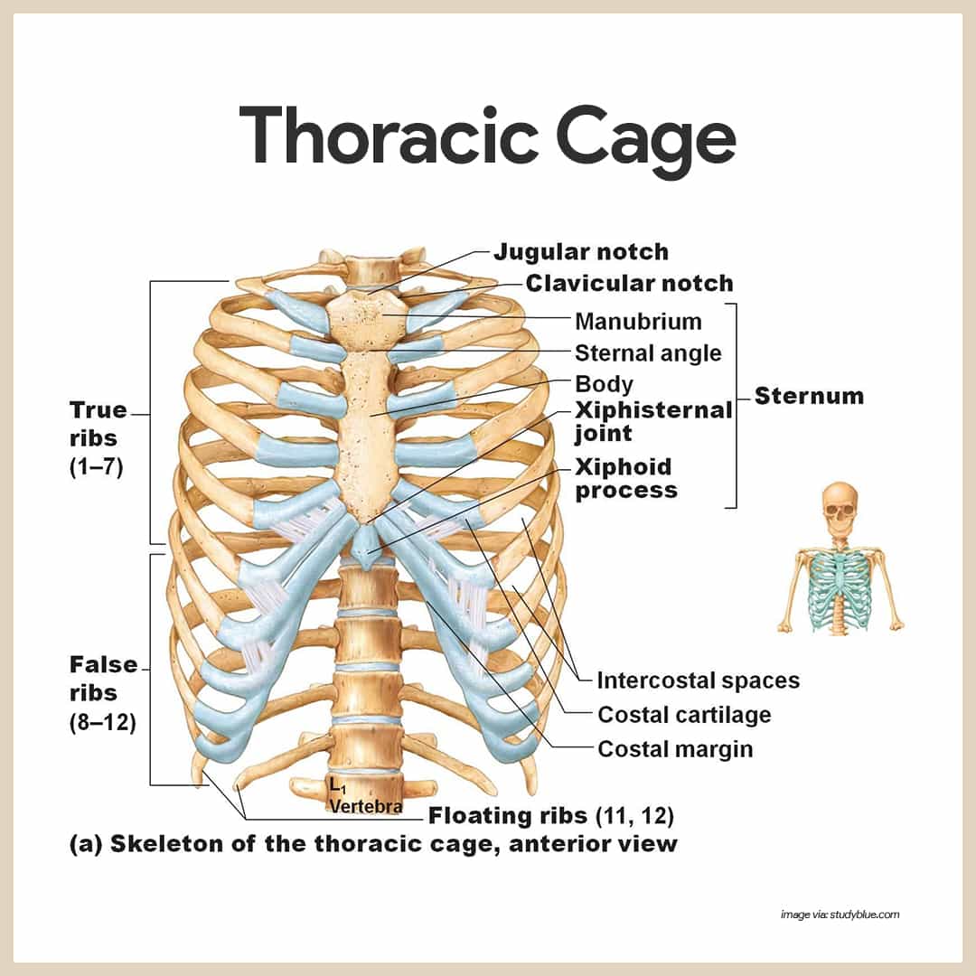
True Ribs (Ribs 1–7): These are also called vertebrosternal ribs. Their name tells you their connection: from a vertebra to the sternum. Each true rib has its own piece of costal cartilage that attaches it directly to the sternum.
-
False Ribs (Ribs 8–12): These are called vertebrochondral ribs. They do not have a direct cartilaginous connection to the sternum. Instead, the costal cartilages of ribs 8, 9, and 10 connect to the cartilage of the rib directly above them, ultimately piggybacking on the cartilage of rib 7.
-
Floating Ribs (Ribs 11–12): These are a subset of the false ribs, sometimes called vertebral ribs. They are the shortest ribs and do not connect to the sternum at all. Their small costal cartilages end within the muscles of the lateral abdominal wall.
The diagram below provides a clear visual of these three categories and the overall structure of the thoracic cage.
Conclusion
In this lesson, we have assembled the final pieces of the axial skeleton. By combining the sternum, ribs, and thoracic vertebrae, we form the thoracic cage, a structure uniquely built for both protection and flexibility.
Key Takeaways:
- The thoracic cage consists of the sternum, 12 pairs of ribs with their costal cartilages, and the 12 thoracic vertebrae.
- The sternum is composed of the superior manubrium, the central body, and the inferior xiphoid process. The sternal angle is a critical palpable landmark for locating the second rib.
- Ribs are classified based on their anterior attachment to the sternum:
- True ribs (1–7) attach directly via their own costal cartilage.
- False ribs (8–12) have an indirect attachment or no attachment.
- Floating ribs (11–12) are a subset of false ribs that do not attach to the sternum.
Having completed our study of the axial skeleton, we are now ready to move outward. In our next lesson, we will begin exploring the appendicular skeleton, starting with the pectoral girdle—the clavicle and scapula—which connects your upper limbs to the axial skeleton at the sternum.