Welcome to our next lesson. In the previous session, we established the foundation of the upper limb by examining the pectoral girdle. You learned how the clavicle and scapula anchor the arm to the axial skeleton, culminating in the glenoid cavity of the scapula—the shallow socket for the shoulder joint.
Today, we will explore the bone that fits into that socket: the humerus. As the single bone of the arm (the region between the shoulder and elbow), the humerus is a classic long bone. Your goal for this lesson is to identify the humerus and its key bony landmarks. A solid understanding of these landmarks is essential for your nursing practice, as they are crucial for understanding arm movements, locating major nerves and blood vessels, and assessing common injuries like fractures.
The Humerus: An Overview
The humerus is the longest and largest bone of the upper limb. Like other long bones, it's best to study it by dividing it into three regions:
- Proximal End: Articulates with the scapula at the shoulder.
- Shaft: The long, central part of the bone.
- Distal End: Articulates with the forearm bones (radius and ulna) at the elbow.
Let's begin with a video that systematically walks through these regions. The presenter, a fellow nurse, does an excellent job explaining why each bump and groove on the bone exists.
Humerus Bone Anatomy and Physiology Lecture
This video, "Humerus Bone Anatomy and Physiology Lecture" from RegisteredNurseRN, provides a clear, landmark-by-landmark tour of the humerus. We will use it as our primary guide.
First, watch the section covering the proximal end, from the start of the anatomy tour. Focus on the parts that form the shoulder joint and provide attachment sites for the powerful shoulder muscles.
Proximal End: The Shoulder Articulation
As you saw in the video, the proximal end of the humerus is dominated by the structures that form the glenohumeral (shoulder) joint and anchor the rotator cuff muscles.
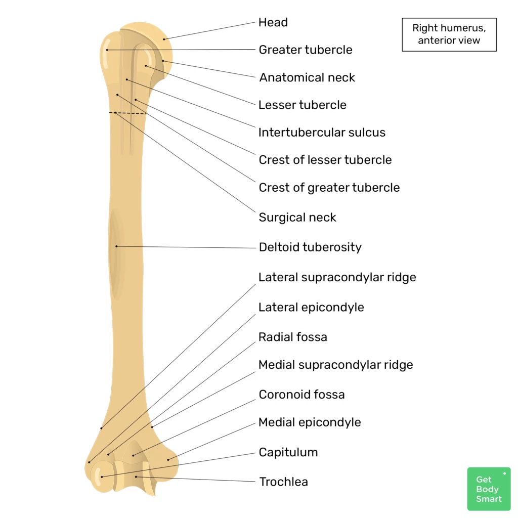
Let's review these key features:
- Head: The large, smooth, rounded structure that articulates with the glenoid cavity of the scapula. This "ball-and-socket" arrangement is what gives the shoulder its incredible mobility.
- Anatomical Neck: A slight groove just below the head, marking the former site of the epiphyseal plate. The fibrous capsule of the shoulder joint attaches here.
- Greater and Lesser Tubercles: These are two prominent bumps just distal to the head. The larger greater tubercle is positioned laterally, while the smaller lesser tubercle is on the anterior side. These serve as critical attachment points for the four rotator cuff muscles you'll learn about later.
- Intertubercular Sulcus (or Groove): The groove located between the two tubercles. It is sometimes called the bicipital groove because the tendon of the long head of the biceps brachii muscle runs through it.
- Surgical Neck: A constriction below the tubercles where the head tapers to meet the shaft. It is named the "surgical" neck because it is a much more common site for fractures than the anatomical neck.
Fractures of the surgical neck are clinically significant because of the important structures that pass nearby. To appreciate this, let's watch a brief clip from another excellent resource.
This video from The Noted Anatomist provides excellent clinical context for the anatomical structures.
Please watch this short segment on the surgical neck, from anatomical vs. surgical neck. Pay close attention to the nerve mentioned and why a fracture here could be problematic.
As the video highlights, the axillary nerve and posterior circumflex humeral artery wrap around the surgical neck. Damage to this nerve from a fracture can lead to paralysis of the deltoid muscle (the main shoulder muscle) and loss of sensation over the shoulder. This is a classic example of how anatomical knowledge directly informs clinical assessment.
The Shaft: A Pathway for Muscles and Nerves
Now let's move down to the shaft, or body, of the humerus. This section primarily serves as an attachment point for various muscles that move the arm.
Humerus Bone Anatomy and Physiology Lecture
Let's return to our main video from RegisteredNurseRN.
Watch the portion covering the shaft, from the radial groove to the supracondylar ridges. Note the key landmark for the deltoid muscle and the groove on the posterior side.
The two most important landmarks on the shaft are:
- Deltoid Tuberosity: A roughened, V-shaped area on the lateral surface of the shaft. This is the insertion point for the deltoid muscle. The powerful pull of this muscle creates this distinctive marking on the bone.
- Radial Groove: A shallow groove that runs diagonally down the posterior surface of the humerus. This groove protects the radial nerve and deep brachial artery as they pass down the arm.
Just like with the surgical neck, the radial groove has important clinical implications. A fracture in the middle of the humeral shaft can damage the radial nerve. This can cause "wrist drop," an inability to extend the wrist and fingers.
Distal End: Forming the Elbow Joint
Finally, we arrive at the distal end of the humerus, which flares out to form the complex structures of the elbow joint. This part articulates with both the ulna and radius of the forearm.
Humerus Bone Anatomy and Physiology Lecture
We'll finish our tour with the RegisteredNurseRN video.
Please watch the final segment covering the distal end of the humerus, from the fossae to the condyles. This section has many new terms, so focus on how the different parts fit together to allow for flexion and extension of the elbow.
The distal end can be broken down into articular and non-articular surfaces.
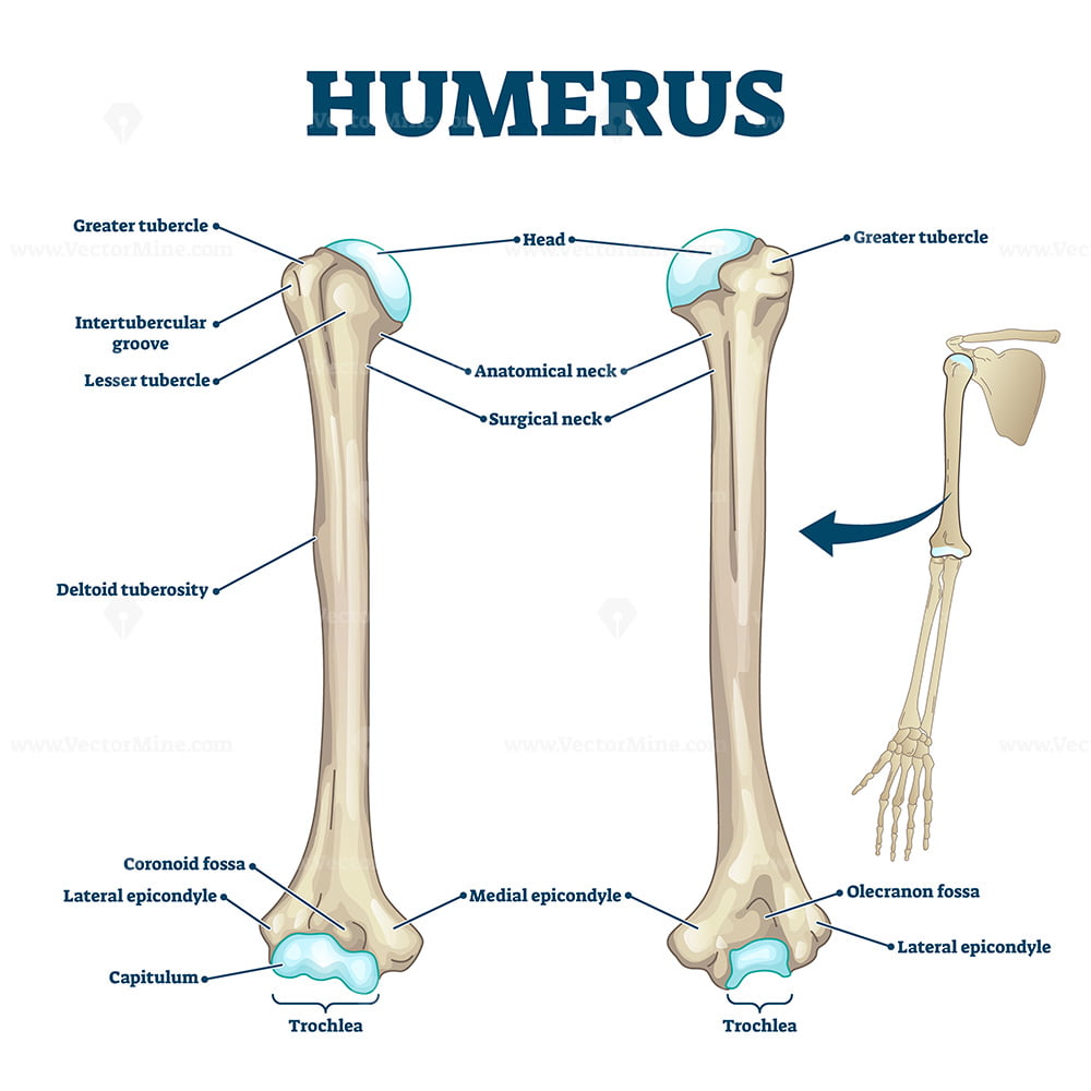
Articular Surfaces (The Condyles):
- Trochlea: A spool-shaped surface on the medial side that articulates with the ulna. Its shape allows for the hinge-like motion of the elbow (flexion and extension).
- Capitulum: A rounded, knob-like surface on the lateral side that articulates with the head of the radius.
Non-Articular Surfaces:
- Medial and Lateral Epicondyles: These are the prominent bumps on either side of the elbow that you can easily feel. They are attachment sites for the powerful muscles of the forearm that flex and extend the wrist and fingers. The medial epicondyle is larger and more prominent.
- Fossae: These are three depressions that accommodate projections from the forearm bones during movement.
- Coronoid Fossa: A depression on the anterior surface, above the trochlea. It receives the coronoid process of the ulna when the elbow is flexed.
- Radial Fossa: A smaller depression on the anterior surface, above the capitulum. It receives the head of the radius when the elbow is fully flexed.
- Olecranon Fossa: A deep depression on the posterior surface. It receives the olecranon of the ulna when the elbow is extended, allowing the arm to straighten fully.
The ulnar nerve runs in a groove on the posterior surface of the medial epicondyle. It is very superficial here, which is why hitting your "funny bone" sends a jarring sensation down your forearm and into your hand—you are actually hitting the ulnar nerve.
To consolidate your understanding of all these landmarks, especially the numerous muscle attachments and clinical relationships, please review the following text.
Humerus: Anatomy and clinical notes
This article from Kenhub provides a comprehensive and well-organized summary of the humerus, including helpful mnemonics.
Please read the sections covering the Proximal end, the Shaft, and the Distal end. The mnemonics provided can be very useful for memorization during your exam preparation.
Conclusion
In this lesson, we have conducted a detailed examination of the humerus, the sole bone of the arm. By breaking it down into its proximal, shaft, and distal regions, you have learned to identify its key landmarks and, crucially, understand their functional and clinical significance.
Key Takeaways:
- The humerus is divided into a proximal end, a shaft, and a distal end.
- The proximal end features the head for articulation with the scapula, the greater and lesser tubercles for muscle attachment, and the clinically important surgical neck.
- The shaft features the deltoid tuberosity for the deltoid muscle and the radial groove which protects the radial nerve.
- The distal end forms the elbow joint with the trochlea (for the ulna) and capitulum (for the radius). It also includes the prominent medial and lateral epicondyles and three fossae (coronoid, radial, olecranon) that accommodate the forearm bones during movement.
- Key fracture sites (surgical neck, mid-shaft, medial epicondyle) are associated with potential damage to specific nerves (axillary, radial, ulnar).
Now that we have established the anatomy of the arm, our next lesson will move to the forearm. We will examine the radius and ulna and see precisely how they articulate with the distal humerus to create the elbow joint.