Hello. Last lesson traced how retained skin cells and sebum can form an open or closed comedone inside a follicle. The practical challenge is that a close-up photograph shows the surface, not the follicle’s contents. A dark dot may be a blackhead, but it may also be a normal sebaceous filament.
This lesson develops a cautious way to compare them: look at shape, texture, size, and distribution together, rather than treating colour as a diagnosis. The examples include different skin tones, because “whitehead” and “blackhead” are names for types of comedones, not reliable descriptions of how every lesion will look against every complexion.
Three appearances, two different processes
An open comedone, or blackhead, is a plug within a follicle whose exposed surface looks dark. Visually, it may appear as a distinct dark centre in a widened opening, sometimes with a slightly raised edge. A closed comedone is also a plugged follicle, but its opening is covered. It tends to appear as a small, raised bump without an exposed dark centre.
A sebaceous filament is different: it is a normal structure associated with the passage of sebum through a follicle, not an acne plug. When visible, filaments often appear as many fine dots of similar size, particularly across the nose. They can be gray, tan, yellowish, or brown. Their colour and visibility vary with the skin and the lighting.
Read Cleveland Clinic’s Sebaceous Filaments: Difference From Blackheads & Treatment for the distinction between a plug and a filament. The comparison is more useful than any proposed “test” involving squeezing; do not squeeze skin to identify what a dot is.
Sebaceous Filaments: Difference From Blackheads & Treatment
Cleveland Clinic explains why a visible follicle is not necessarily a blocked one, then describes where sebaceous filaments are commonly noticeable.
Under “Overview,” find “What’s the difference between sebaceous filaments and blackheads?” Read from the paragraph beginning “Blackheads are a type of acne” through the plug distinction. Continue through the paragraph beginning “Blackheads look like a dark speck” and ending with the filament colours the appearance comparison. Then, under “How common are sebaceous filaments?”, read from “Every person has sebaceous filaments” to the end of the paragraph listing where they appear the distribution clues. Focus on plug versus normal oil passage and on patterns across multiple follicles. The later discussion of what comes out when skin is squeezed is not an identification method to try.
One useful visual rule follows from this distinction: a pattern tells you more than an isolated dot. A field of tiny, similar-looking dots in many neighbouring nasal pores favours visible sebaceous filaments. A more conspicuous, individually distinguishable dark centre in a widened opening makes a blackhead more plausible. Neither rule is absolute: blackheads can occur in groups, and a photograph may exaggerate the apparent size of pores.
This short segment from Dr Dray describes why filaments are especially easy to mistake for blackheads on the nose.
Blackheads on the Nose? Here’s What They Really Are | Derm Explains
In “Blackheads on the Nose? Here’s What They Really Are,” dermatologist Dr Dray describes sebaceous filaments as normal structures rather than acne plugs.
Watch sebaceous filaments, beginning when the discussion turns from blackheads to filaments and ending before the next look-alike is introduced. Concentrate on their fine appearance and tendency to become visible again. The point is not that washing can permanently remove a normal structure.
Annotated example: fine dots across a nose
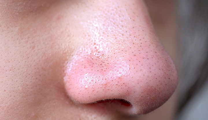
Read this image in layers. First, distribution: the dots recur across much of the nose rather than presenting as one or two standout lesions. Next, relative size: most are small compared with the surrounding visible pore pattern. Finally, surface shape: the image does not clearly show a separate raised bump around each dot. Together, these observations make sebaceous filaments a reasonable interpretation of the overall pattern.
That interpretation does not mean every dot in the photograph must be a filament. A person can have filaments and blackheads in the same area. Nor does the dark colour prove there is a plug: the camera is recording tiny openings under particular lighting, not examining their contents.
A true blackhead on a nose would be more plausible if one opening stood out as larger or more distinctly plugged than its neighbours, with a focal dark centre. “More plausible” is the appropriate level of confidence. Magnification, shadow, and skin texture can all change how large or dark an opening appears.
Annotated example: bumps without dark centres
Closed comedones require a change of emphasis. Instead of looking for a dark point, look for a small elevation of the skin. Several may create an uneven, finely bumpy surface on the forehead or cheeks. Their colour can be close to the surrounding skin; they need not look white.
Dermatologist Dr Aleksandra Brown describes this surface appearance in the opening of How to Get Rid of Closed Comedones.
How to Get Rid of Closed Comedones | Dermatologist Explains Whiteheads & Bumpy Skin
This segment provides a visual and tactile description of small, covered follicular bumps, complementing the pore cross-section from the previous lesson.
Watch closed comedones, from the definition through the examples of where the bumps commonly appear. Focus on the raised, sometimes finely uneven texture—not on assuming that every small bump must be acne.

Here, the arrows direct attention to raised lesions, not to dark material at open pores. The surrounding forehead also has multiple small bumps. That combination supports “likely closed comedones” more than “sebaceous filaments”: filaments usually present as fine dots within visible openings, especially on the nose, rather than as a collection of covered elevations.
The photographed skin is medium-brown, and some bumps look lighter than the surrounding area while others are closer to its colour. On darker or lighter skin, the contrast can differ again. The dependable clue is the bump with no exposed dark centre, not whether it satisfies the everyday word “white.”
Applying the same clues across skin tones
Colour words can mislead when used alone. A filament may look light gray against one complexion and brown against another. A blackhead’s exposed centre may look dark, but the contrast between that centre and nearby skin varies. A closed comedone may be pale, skin-coloured, or simply more noticeable for the change in texture it creates.
Consider three constructed visual descriptions, not photographs or diagnoses:
- Lighter skin, many nasal dots: Most openings show similarly sized gray-tan points lying nearly flat. The repetition and lack of standout bumps favour visible sebaceous filaments over dozens of separate blackheads.
- Deep-brown skin, a few standout openings: Among many fine, fairly uniform nasal dots, two openings have noticeably larger, sharply defined dark centres. The background pattern may represent filaments; the two distinctive openings make blackheads possible. Colour alone would not settle either classification.
- Brown skin, a bumpy forehead: Numerous small elevations are close to the surrounding skin colour and have no obvious dark centre. Their raised, clustered appearance makes closed comedones plausible, even if none looks white.
These examples use the same reasoning on each skin tone. Compare a feature with the nearby skin and neighbouring follicles, not with a stock image or a fixed colour chart. Check whether it is flat or raised, whether it has a distinct exposed centre, and whether it is one conspicuous lesion or part of a uniform field.
Even then, stop short of certainty. A flat dark mark is not automatically a blackhead, and a small bump is not automatically a closed comedone. The next lesson examines several look-alikes and the point at which an appearance is too uncertain for self-classification.
What a photograph can—and cannot—tell you
A photo can support an appearance-based description: “fine, uniform dots across the nose” or “multiple small raised bumps without dark centres.” It usually cannot show the follicular plug directly. It also leaves out useful context, such as how the surface feels, whether a feature is changing, and how lighting or image processing has altered its appearance.
Use these distinctions as a guide to likelihood, not as a reason to press, pick, or use a tool for confirmation. Removal is particularly unhelpful as a diagnostic idea for sebaceous filaments: they are normal structures and can become visible again. Later lessons will address the risks of extraction and what noninvasive care can realistically achieve.
Key takeaways
A likely blackhead has a distinct dark centre associated with a plugged, open follicle. A likely closed comedone is a small raised bump without that exposed centre. Visible sebaceous filaments commonly form a fine, fairly uniform pattern of dots, especially on the nose, and are not acne plugs. Shape and distribution help more than colour, but none of these visual clues makes a photograph a definitive diagnosis.
Next, you will compare these patterns with other small bumps and dark spots that may resemble comedones—and learn when the sensible conclusion is simply that the appearance needs professional assessment.