Introduction
Welcome to Module 5: Back Anatomy and Hypertrophy Application. In the previous module, we concluded our deep dive into the chest by learning how to translate anatomical knowledge into precise coaching cues. We are now shifting our focus from the front of the torso to the back—a complex and powerful network of muscles responsible for everything from creating a "V-taper" aesthetic to generating immense pulling strength.
This lesson begins our exploration of the back with its largest and most prominent muscle: the latissimus dorsi. Your learning outcome is to describe the origin, insertion, and fiber direction of the latissimus dorsi and identify its primary joint actions.
Mastering the anatomy of the "lats" is fundamental. Because of its vast size and multiple attachment points, different parts of the muscle are best targeted by different movements. Understanding this foundational architecture is the first step toward building a truly effective back training program.
1. The "Broadest Muscle of the Back"
The name latissimus dorsi literally translates from Latin to "broadest muscle of the back," a fitting title for the largest muscle in the upper body by surface area. It's the primary muscle that gives the back its width.
Before we break down its components, let's get a visual overview of this impressive muscle.
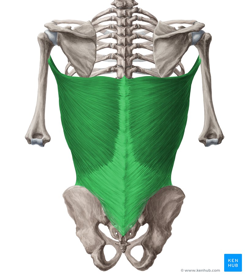
2. Origin: A Vast, Fan-Shaped Foundation
The latissimus dorsi has an incredibly broad origin, meaning it attaches to a wide array of structures on the posterior torso. Think of these as the anchor points from which the muscle will pull.
To visualize these attachments, please watch the following video segment.
The Latissimus Dorsi: Anatomy and function
The video 'The Latissimus Dorsi: Anatomy and function' from Muscle & Motion provides excellent 3D animations of the lat's origin points.
Watch from 00:18 to 01:06. Pay close attention to how the muscle attaches to the spine, the pelvis, and the ribs.
As you saw, the latissimus dorsi originates from:
- Spinous processes of the thoracic and lumbar vertebrae (T7-L5): The bony bumps you can feel running down the center of your mid-to-lower back.
- Thoracolumbar Fascia: A thick sheet of connective tissue that covers the deep muscles of the lower back, connecting the lats to the sacrum and iliac crest.
- Posterior Iliac Crest: The back ridge of your pelvic bone.
- Lower 3-4 Ribs: It interdigitates (interlocks like fingers) with the external oblique muscles here.
- Inferior Angle of the Scapula: The bottom tip of the shoulder blade (this attachment is present in about half the population).

The key takeaway here is that the lats connect the arm directly to the spine and pelvis, giving it immense leverage to pull the arm towards the body.
3. Insertion and Fiber Twist: Convergence to a Point
While the origin is broad and distributed, all of these muscle fibers converge, run upwards and outwards, and insert at a single, narrow point.
This video from AnatomyZone provides a clear explanation of the insertion and a crucial detail about how the fibers arrange themselves.
Latissimus Dorsi | Muscle Anatomy
This segment clearly shows where the latissimus dorsi attaches to the arm bone (humerus) and explains the 'twist' in the muscle fibers.
Watch from 01:43 to 02:26. Focus on two key points: the location of the insertion on the humerus (intertubercular sulcus) and the description of how the fibers 'twist' before they attach.
-
Insertion: The latissimus dorsi inserts on the floor of the intertubercular sulcus of the humerus. This is a groove on the front of your upper arm bone. The fact that a back muscle attaches to the front of the arm is a critical detail that explains one of its key functions.
-
The Anatomical Twist: As the video explained, the fibers twist on themselves before inserting.
- The highest fibers from the thoracic spine wrap around and insert on the lowest part of the tendon.
- The lowest fibers from the pelvis and iliac crest run up and insert on the highest part of the tendon.
This twist is not just a random quirk; it has significant biomechanical implications that we will explore when we discuss exercise selection.
4. Fiber Direction: Three Functional Divisions
Because the lats originate from such a wide area and converge on a single point, their fibers run in different directions. We can conceptually group these fibers into three functional divisions based on their orientation. This is similar to how we analyzed the different divisions of the pectoralis major.
You can think of these divisions as specialized sub-routines in a larger program. While they all contribute to the main goal of pulling the arm, each is optimized for a slightly different line of pull.
The Ultimate Guide to Lat Pulldowns
The article 'The Ultimate Guide to Lat Pulldowns' by Conor Harris does an excellent job of breaking down the lats into these functional divisions.
Read the three short sections titled 'Thoracic Fibers', 'Lumbar Fibers', and 'Iliac Fibers'. As you read, notice how the fiber orientation (horizontal, diagonal, vertical) is described for each division.
To summarize the fiber directions:
- Thoracic (Upper) Fibers: Run mostly horizontally.
- Lumbar (Middle) Fibers: Run diagonally upwards.
- Iliac (Lower) Fibers: Run almost vertically upwards.
Understanding these different lines of pull is the key to selecting exercises that target the entire latissimus dorsi muscle, not just one portion of it. For example, a horizontal pull (like a row) will better align with the thoracic fibers, while a vertical pull (like a pulldown) will better align with the iliac fibers.
5. Primary Joint Actions
Now, let's put it all together. A muscle creates movement by pulling its insertion point towards its origin point. Given the lat's attachments, what happens when it contracts?
This final video clip provides a clear, animated demonstration of the lat's primary functions at the shoulder (glenohumeral) joint.
The Latissimus Dorsi: Anatomy and function
Watch this brief segment from Muscle & Motion to see the lat's main actions animated.
Watch from 01:25 to 02:00. The video labels the three primary actions clearly. Try to mimic each movement with your own arm.
The primary joint actions of the latissimus dorsi are:
- Shoulder Extension: Pulling the arm down and back from a position in front of the body. Think of the motion in a dumbbell row or a straight-arm pulldown.
- Shoulder Adduction: Pulling the arm down towards the side of the body from an out-to-the-side position. This is the primary action in a lat pulldown or a pull-up.
- Shoulder Internal (Medial) Rotation: Turning the arm inward. This action is due to the muscle's attachment on the front of the humerus. It works with the pecs to create powerful internal rotation.
All "lat" exercises in the gym are simply variations and combinations of these three fundamental movements.
Conclusion
In this lesson, you've dissected the foundational anatomy of the latissimus dorsi. You now have the anatomical blueprint needed to understand how this crucial muscle functions.
Key Takeaways:
- Origin: The lats have a vast, fan-shaped origin on the spine (T7-L5), pelvis (iliac crest), and ribs.
- Insertion: All fibers converge and twist to insert onto a small point on the front of the humerus (intertubercular sulcus).
- Fiber Direction: The fibers have distinct orientations: the upper fibers are more horizontal, the middle are diagonal, and the lower are more vertical.
- Primary Actions: The lats are powerful shoulder extensors, adductors, and internal rotators.
Preview for the Next Lesson
While the lats are dominant, they don't work alone. In our next lesson, we will move to the muscles of the mid and upper back: the trapezius and the rhomboids. We will explore their distinct anatomy and how their primary function—controlling the scapula—differs from and complements the function of the lats.