Introduction
Welcome to Module 9. In our previous lessons, we completed an in-depth analysis of the quadriceps, moving from anatomical structure to identifying and correcting common training faults with precise coaching cues. We have effectively covered the anterior (front) aspect of the upper leg.
Now, we pivot to the posterior chain, a critical driver of athletic power and physique development. This module will focus on the glutes and hamstrings. Today, we begin with the anatomical foundation of the gluteal group. Our learning outcome is to describe the origin, insertion, and fiber direction of the gluteus maximus, medius, and minimus.
Understanding this core anatomy is like learning the syntax of a programming language. Before you can write effective code (select exercises), you must understand the rules and structure of the components you're working with. The origin, insertion, and fiber direction of a muscle dictate its "line of pull" and, therefore, its function.
The Gluteal Complex: A Layered System
The muscles colloquially known as "the glutes" are actually a complex of three distinct muscles arranged in layers. From superficial (closest to the skin) to deep, they are:
- Gluteus Maximus
- Gluteus Medius
- Gluteus Minimus
This layered arrangement is crucial. The larger, more superficial muscles often produce powerful, large-scale movements, while the deeper muscles play a key role in stabilization and finer control.
Let's begin by visualizing their layout.
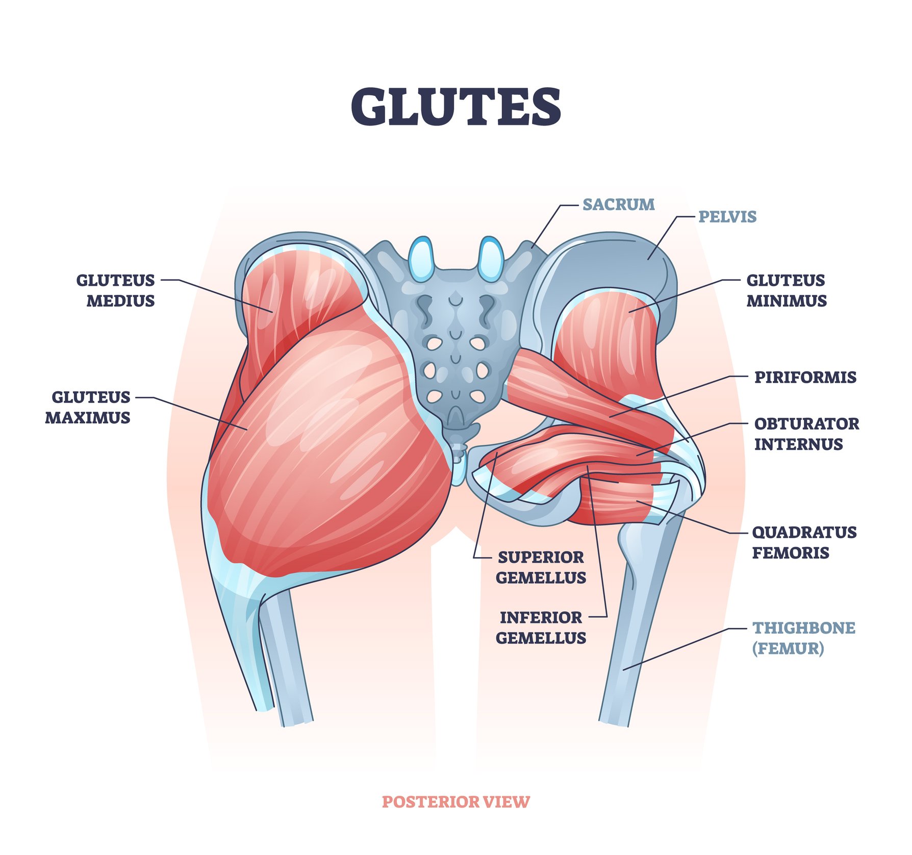
We will now break down each muscle individually.
1. Gluteus Maximus: The Powerhouse
The gluteus maximus is the largest and most powerful muscle in the human body. Its size and strength are defining features of our bipedal locomotion, enabling us to stand, run, and climb.
To understand its structure, let's watch a short, focused video.
The Glutes│Anatomy Tips, Tricks and Mnemonics
This video from Memorize Medical provides a clear and concise overview of the gluteus maximus's attachments and fiber orientation.
Watch from 00:09 to 01:37. Pay close attention to: The origin points on the posterior ilium, sacrum, and coccyx (the central bones of the lower back and tailbone). The two distinct insertion points: the IT band and the gluteal tuberosity of the femur. The diagonal direction of the muscle fibers.
Let's formalize those details:
-
Origin: The gluteus maximus originates from a broad area along the posterior (back) midline of the body.
- Posterior surface of the ilium (the large, wing-like pelvic bone).
- Dorsal (back) surface of the sacrum (the triangular bone at the base of the spine).
- Lateral (side) aspect of the coccyx (the tailbone).
-
Insertion: From this wide origin, the fibers converge and insert onto two main points on the upper thigh bone (femur).
- The upper, larger portion inserts into the iliotibial (IT) band, a thick sheath of connective tissue running down the outside of the thigh.
- The lower, deeper portion inserts onto the gluteal tuberosity, a roughened patch on the back of the femur.
-
Fiber Direction: Critically, the fibers of the gluteus maximus are not vertical. They run diagonally downward and outward, at approximately a 40-45 degree angle, from the spine and pelvis toward the side of the thigh. This diagonal orientation is the key to its function, allowing it to pull the thigh bone backward and rotate it outward.
2. Gluteus Medius & Minimus: The Upper Shelf and Deep Stabilizer
Lying partially underneath the gluteus maximus are the gluteus medius and minimus. They sit on the upper, outer aspect of the hip, forming what is often called the "upper glute shelf." They are primary stabilizers of the pelvis and are crucial for hip abduction (moving the leg out to the side).
Let's watch another brief clip to locate these muscles.
The Glutes│Anatomy Tips, Tricks and Mnemonics
The same video now covers the gluteus medius and minimus, highlighting their relationship to each other and their shared insertion point.
Watch from 01:49 to 02:23. Note how the gluteus medius has a larger 'footprint' on the ilium than the maximus, and how the minimus lies directly beneath it.
Now, we'll examine their individual anatomy.
Gluteus Medius
- Origin: It originates from the outer surface of the ilium, between the iliac crest (the top ridge of your hip bone that you can feel with your hands) and the posterior gluteal line.
- Insertion: The fibers converge and insert onto the lateral surface of the greater trochanter of the femur. This is the prominent bony knob you can feel on the outside of your hip.
- Fiber Direction: The gluteus medius has a distinct fan-shaped fiber orientation. As the image below illustrates, it's not a single sheet of parallel fibers. It has anterior (front), middle, and posterior (back) fibers, all converging on the same point. This multi-directional pull allows it to perform several actions, which we will explore in the next lesson.

Gluteus Minimus
- Origin: It originates from the outer surface of the ilium, directly underneath the gluteus medius.
- Insertion: Like the medius, its fibers converge to insert on the greater trochanter of the femur, but slightly more toward the front (anteriorly).
- Fiber Direction: It is also fan-shaped, mirroring the gluteus medius that it lies beneath. Because they are so similar in location and fiber orientation, they work together as primary hip abductors and stabilizers.
Summary and Key Anatomical Relationships
Having a clear mental map of these muscles is crucial. Let's consolidate this information into a reference table.
| Muscle | Origin | Insertion | Fiber Direction |
|---|---|---|---|
| Gluteus Maximus | Posterior ilium, sacrum, coccyx | Gluteal tuberosity (femur) & Iliotibial (IT) band | Diagonal (downward and outward) |
| Gluteus Medius | Outer surface of the ilium | Lateral surface of the greater trochanter (femur) | Fan-shaped (anterior, middle, posterior fibers) |
| Gluteus Minimus | Outer surface of the ilium (deep to the gluteus medius) | Anterior surface of the greater trochanter (femur) | Fan-shaped (mirrors gluteus medius) |
To help solidify this knowledge, I recommend you read through this short, clear reference guide. It reinforces the exact points we've covered.
Glute Muscles Guide: Anatomy, Structure & Biological Function
This guide from Booty Builder provides a clean, text-based summary of the origin, insertion, and structure of all three gluteal muscles.
Read the sections titled 'Gluteus Maximus', 'Gluteus Medius', and 'Gluteus Minimus'. Focus on how the descriptions of origin, insertion, and structural characteristics match what you've learned in this lesson.
Coach's Note: Why Fiber Direction is Everything
As a coach, the concept of "line of pull" is your north star for exercise selection. A muscle can only contract and pull its insertion point closer to its origin point.
- Because the gluteus maximus fibers run diagonally from the midline to the outer thigh, they primarily pull the thigh backward (hip extension).
- Because the gluteus medius/minimus fibers run from the outer hip bone down to the side of the thigh, they primarily pull the thigh outward, away from the body's midline (hip abduction).
This is why an exercise like a hip thrust (hip extension) primarily targets the gluteus maximus, while an exercise like a side-lying leg raise (hip abduction) targets the gluteus medius/minimus. Their anatomical structure dictates their function.
Conclusion
In this lesson, we have established the foundational anatomy of the gluteal complex. You can now describe the origin, insertion, and fiber direction for the gluteus maximus, medius, and minimus.
Key Takeaways:
- The glutes are a layered system of three muscles: maximus (superficial), medius (intermediate), and minimus (deep).
- Gluteus Maximus: Originates from the back of the pelvis/sacrum and inserts on the back/side of the femur and IT band, with diagonal fibers designed for power.
- Gluteus Medius & Minimus: Originate from the outer "shelf" of the hip bone and insert on the bony knob on the side of the hip (greater trochanter), with fan-shaped fibers designed for abduction and stability.
- A muscle's structure—specifically its fiber direction—determines its function.
Preview of the Next Lesson
Now that we have built our anatomical map, we will bring it to life. In our next lesson, we will differentiate the primary roles of the gluteus maximus (hip extension) from the medius/minimus (hip abduction, stabilization). We will explore their specific joint actions and begin to connect this knowledge to the movements you see every day in the gym.