Introduction
Welcome to the third lesson in our module on the glutes and hamstrings. In the previous lesson, we established the distinct functional roles of the gluteal muscles, highlighting the gluteus maximus as the primary hip extensor and the gluteus medius/minimus as crucial stabilizers and abductors.
Today, we shift our focus slightly lower down the posterior chain to the hamstrings. These muscles are powerful synergists to the glutes in hip extension and are the primary movers for knee flexion. A detailed understanding of their anatomy is non-negotiable for designing a complete leg development program.
Our learning outcome for this lesson is to describe the origin, insertion, and fiber direction of the hamstring muscles: biceps femoris, semitendinosus, and semimembranosus. By dissecting their individual attachments, we will lay the groundwork for understanding why certain exercises, like Romanian deadlifts, feel different from exercises like leg curls.
Meet the Hamstrings
The hamstrings are not a single muscle but a group of muscles located on the posterior (back) of your thigh. They run from your pelvis down to your lower leg bones. Traditionally, this group consists of three muscles, but from a functional and training perspective, it's more accurate to think of them as four distinct muscle bellies.
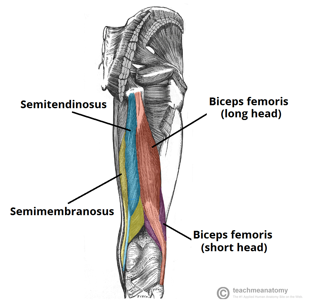
The three main muscles are:
- Biceps Femoris (on the lateral side, or outside of the thigh)
- Semitendinosus (more medial, in the middle of the group)
- Semimembranosus (the most medial, or innermost of the group)
The Biceps Femoris is further divided into a long head and a short head, giving us our four functional muscle bellies.
Let's begin with a quick video overview that introduces these muscles and their general attachments.
The Most Effective Way to Train HAMSTRINGS | Training Science Explained
This video from Jeff Nippard provides a concise, training-focused introduction to the four hamstring muscles and their basic attachments.
Watch from 00:35 to 01:23. Pay close attention to the names of the four muscles, their locations (medial vs. lateral), and which bones they attach to (pelvis, tibia, fibula). Notice the key distinction made about the Biceps Femoris short head.
The Common Origin: The Ischial Tuberosity
As the video mentioned, three of the four hamstring muscles share a common origin point: the ischial tuberosity. This is a bony prominence at the bottom of your pelvis. You can feel it if you sit on a hard surface and rock from side to side; it's often called the "sitting bone."
This shared origin is a crucial piece of the puzzle. It means that to stretch these three muscles at the hip, you must tilt your pelvis forward, which is the foundational mechanic of a hip hinge movement like a Romanian Deadlift (RDL).
The following video provides a more clinical and detailed look at these attachments.
Clinical Anatomy of the Hamstrings | Expert Physio Review
The Clinical Physio channel gives a precise anatomical tour of the hamstrings. This first segment clearly identifies the common origin point.
Watch from 01:10 to 01:47. The key takeaway here is the visualization of the ischial tuberosity as the common anchor point for the semimembranosus, semitendinosus, and the long head of the biceps femoris.
The Anatomical Blueprint: Individual Attachments
Now that we have the general layout, let's build a precise blueprint of each muscle's origin and insertion. Understanding these exact points is what allows you to visualize the muscle's line of pull and, therefore, its function.

To get the specific details, we'll consult a detailed text resource.
Physiopedia is an excellent reference for precise anatomical descriptions. We will use it to break down the origin and insertion of each component of the hamstring group.
Read the following sections under the 'Anatomy' heading: Semimembranosus: Note its origin on the ischial tuberosity and insertion on the medial tibial condyle. Semitendinosus: Note its origin (also on the ischial tuberosity) and its insertion on the medial surface of the tibia. Biceps femoris - Long head: Note its origin on the ischial tuberosity and insertion on the head of the fibula (the lateral lower leg bone). Biceps femoris – Short head: Pay special attention here. Note its origin is not on the pelvis, but on the femur itself (linea aspera). It then joins the long head to insert on the head of the fibula.
Let's consolidate this information into a table for easy reference. This is your anatomical cheat sheet for the hamstrings.
| Muscle | Origin | Insertion | Location |
|---|---|---|---|
| Semimembranosus | Ischial Tuberosity (sitting bone) | Medial Condyle of Tibia (inner shin bone) | Medial |
| Semitendinosus | Ischial Tuberosity (sitting bone) | Medial Surface of Tibia (inner shin bone) | Medial |
| Biceps Femoris (Long Head) | Ischial Tuberosity (sitting bone) | Head of Fibula (outer shin bone) | Lateral |
| Biceps Femoris (Short Head) | Linea Aspera of Femur (back of thigh bone) | Head of Fibula (outer shin bone) | Lateral |
Fiber Direction and Muscle Architecture
A muscle's fiber direction is simply the imaginary line connecting its origin to its insertion. This line of pull dictates the muscle's primary action.
- Medial Hamstrings (Semimembranosus & Semitendinosus): Their fibers run almost vertically down the posterior-medial aspect of the thigh. This path is ideal for pulling the tibia straight back (knee flexion) and pulling the pelvis down/back (hip extension).
- Lateral Hamstrings (Biceps Femoris): Their fibers run diagonally from the relatively medial ischial tuberosity down and out to the lateral fibula. This postero-lateral path is also great for knee flexion and hip extension, but the diagonal pull gives it an additional rotational component.
Beyond simple direction, the internal architecture of the fibers matters. This is a concept that should resonate with your background in optimizing complex systems. Muscles exhibit trade-offs between force production and range of motion, largely determined by their fiber arrangement (pennation).
Hamstring - exerciseprescriptor
The Exerciseprescriptor resource offers a fantastic explanation of muscle fiber arrangement in the hamstrings, which directly relates to their function and injury risk.
Read the section titled 'Muscle fiber arrangement and force production.' Focus on: The definitions of pennate and parallel fibers. The functional trade-off: Pennate fibers allow for more force, while parallel fibers allow for greater range of motion and contraction speed. The specific arrangement for each hamstring muscle and its practical implication, especially regarding injury risk for the biceps femoris.
This concept explains why the biceps femoris, with its pennated, force-oriented but less extensible fibers, is the most commonly injured hamstring muscle in high-speed activities like sprinting. Its architecture prioritizes force production over rapid lengthening, making it vulnerable.
Coach's Note: The "Fourth Hamstring"
You may occasionally hear coaches or anatomists refer to the "ischial head of the adductor magnus" as the "fourth hamstring." This is because, like the main hamstrings, this part of the adductor magnus muscle originates on the ischial tuberosity and assists in hip extension. While technically an adductor, it functions very similarly to a hamstring in hip-extension movements like squats and RDLs. This is an advanced detail, but it's useful for understanding the deep connections within the posterior chain.
Conclusion
In this lesson, we have constructed a detailed anatomical map of the hamstring muscle group.
Key Takeaways:
- The hamstrings consist of three muscles with four functional heads: Semimembranosus and Semitendinosus (medial), and the Biceps Femoris (long and short heads) (lateral).
- The three main, bi-articular hamstrings share a common origin on the ischial tuberosity. This is a critical point for understanding how to stretch them at the hip.
- The short head of the biceps femoris is unique; it originates on the femur and only crosses the knee joint.
- The hamstrings insert on the lower leg bones: the medial hamstrings on the tibia and the lateral hamstrings on the fibula.
- Fiber direction dictates function, with the architecture (pennate vs. parallel) creating a trade-off between force production and excursion range.
Preview of the Next Lesson
We have now laid the complete anatomical foundation. In our next lesson, we will use this knowledge to explore a pivotal concept for training: the biarticular nature of the hamstrings. We will explain how this property affects exercise selection and why you need different movements to fully train the hamstrings in their lengthened position at both the hip and the knee.