Introduction
Welcome to Module 6, where we shift our focus to the shoulder complex. In our last lesson, you learned how to apply specific, anatomy-based coaching cues to correct common back training errors. We established that a deep understanding of scapular mechanics is crucial for effective pulling movements. That knowledge will serve as a direct foundation for this module, as the scapula is the platform upon which the shoulder operates.
This lesson begins our deep dive into the deltoid muscle, the primary muscle responsible for the shoulder's rounded, powerful appearance. Our goal is to describe the origin, insertion, and fiber direction of the anterior, lateral, and posterior deltoid heads. Understanding these anatomical properties is the first and most critical step in designing effective shoulder training programs, as it dictates how each part of the muscle functions.
1. The Deltoid: A Multipennate Powerhouse
The deltoid gets its name from the Greek letter delta (Δ) due to its classic triangular shape. It's a thick, powerful muscle that drapes over the glenohumeral (shoulder) joint.
From a structural standpoint, the deltoid is a multipennate muscle. This means its fibers (fascicles) are arranged at an angle to the tendon, similar to how barbs are arranged on a feather's quill. This architecture allows for more muscle fibers to be packed into a smaller area, leading to greater force production capacity.

For training purposes, we divide the deltoid into three distinct heads, each with a unique origin point and fiber orientation:
- Anterior Deltoid (Front)
- Lateral Deltoid (Side or Middle)
- Posterior Deltoid (Rear)
2. The Common Ground: The Deltoid's Insertion
Before we explore what makes each deltoid head unique, let's start with their single common feature: the insertion point. All three heads converge into one strong tendon that attaches to the humerus (your upper arm bone).
- Insertion: Deltoid tuberosity on the lateral (outer) side of the humerus.
The deltoid tuberosity is a rough patch of bone located about halfway down the outside of your upper arm. The fact that all fibers pull on this single point is crucial. The direction of that pull, determined by the origin of the contracting fibers, is what dictates the resulting movement of the arm.
This animated video provides an excellent visual overview of the deltoid's structure.
Deltoid Muscle Anatomy: Origin, Insertion, Nerve supply and Actions : Dr G Bhanu Prakash
This video from Dr. G Bhanu Prakash will help you visualize the overall structure. Pay close attention to how the three distinct heads all travel downwards to converge at one single insertion point.
Watch the segment from 03:22 to 04:27. This part clearly shows the fibers running inferiorly (downwards) and inserting into the deltoid tuberosity on the humerus.
3. The Origins and Fiber Directions: A Story of Three Heads
The distinct functions of the three deltoid heads come from their different starting points, or origins, and the resulting direction of their muscle fibers. Think of it like three ropes tied to the same post (the humerus), but anchored at different points around a room. Pulling on each rope will move the post in a different direction.
Let's break down each head's anatomy. The following image provides a clear map of these structures, which we will dissect below.
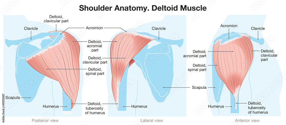
To get a more detailed and dynamic understanding, we'll consult Dr. Prakash's video again.
Deltoid Muscle Anatomy: Origin, Insertion, Nerve supply and Actions : Dr G Bhanu Prakash
This segment provides a clear, animated breakdown of the specific origin point for each of the three deltoid heads.
Watch from 00:27 to 03:22. The video labels the heads as clavicular (anterior), acromial (middle/lateral), and scapular (posterior). Focus on identifying the specific bony landmarks for each origin: the clavicle, the acromion, and the spine of the scapula.
Here is a summary of the origins and fiber directions, which you can cross-reference with the video and image above.
Anterior (Clavicular) Deltoid
- Origin: The anterior (front) surface of the lateral one-third of the clavicle (your collarbone).
- Fiber Direction: The fibers run downward and backward (inferoposteriorly) to the deltoid tuberosity. Imagine fibers starting at the front of your collarbone and angling down and out to the side of your arm.
Lateral (Acromial) Deltoid
- Origin: The lateral (outer) border of the acromion process of the scapula. The acromion is the bony "shelf" at the very top of your shoulder that you can feel with your fingers.
- Fiber Direction: The fibers run almost directly downward in a multipennate fashion to the deltoid tuberosity. This direct line of pull is why this head is the primary muscle for lifting the arm straight out to the side.
Posterior (Spinal) Deltoid
- Origin: The lower lip of the spine of the scapula (the prominent bony ridge on the back of your shoulder blade).
- Fiber Direction: The fibers run downward and forward (inferoanteriorly) to the deltoid tuberosity. Imagine fibers starting from the ridge on your shoulder blade and angling down and forward to the side of your arm.
This table provides a concise summary of the deltoid anatomy.
The website Bodyworks Prime offers a clear and simple table summarizing the origin and insertion points for each deltoid head. It's a great quick-reference tool.
Review the 'Overview' table at the very top of the article. It neatly lists the origin for the anterior, intermediate (lateral), and posterior heads, along with their common insertion.
4. Advanced Concept: The Seven-Segment Model
Your background in computer engineering suggests you appreciate systems with underlying complexity and logic. The three-head model of the deltoid is a functional and effective simplification, much like a high-level API. However, the underlying "code" is more intricate.
Anatomical research reveals the deltoid is actually composed of at least seven distinct segments, defined by their relationship with internal intramuscular tendons.
For a deeper, more technical understanding, this article from Musculoskeletal Key explains the seven-segment model. This level of detail is not typically covered in basic personal training courses but is highly relevant for understanding concepts like regional hypertrophy.
Read the initial section of the article, starting from 'The origin and insertion of the deltoid...' down to the end of the paragraph '...these anatomical segments based on the intramuscular tendons should be taken into consideration when the function of the deltoid is discussed.' Focus on how the classical three heads are further subdivided based on their tendinous attachments.
Why this matters for coaching: This advanced model helps explain why subtle changes in arm path or rotation during an exercise can seem to target slightly different parts of the deltoid. It's the anatomical basis for regional hypertrophy, where one part of a muscle can grow more than another. We will revisit this concept when we discuss exercise selection and biomechanics in later lessons.
Conclusion
In this lesson, you have built the anatomical foundation for understanding the shoulder. You now know the specific origin, insertion, and fiber direction for each of the three functional heads of the deltoid.
Key Takeaways:
- The deltoid is a multipennate muscle composed of the anterior, lateral, and posterior heads.
- All three heads share a common insertion point: the deltoid tuberosity on the humerus.
- Their distinct functions arise from their unique origins:
- Anterior: Lateral clavicle.
- Lateral: Acromion of the scapula.
- Posterior: Spine of the scapula.
- The fiber direction of each head dictates its primary line of pull (e.g., anterior fibers pull forward, lateral fibers pull sideways, posterior fibers pull backward).
- The simplified three-head model can be further broken down into a more complex seven-segment model, which has implications for advanced training concepts.
Preview for the Next Lesson
Now that we know the "what" and "where" of the deltoid's structure, the next logical step is to understand the "how." In our next lesson, we will explore the specific joint actions of each deltoid head and classify their roles as prime movers, synergists, and stabilizers across various common gym movements. This is where anatomy truly begins to inform action.