Hello! Welcome to your next lesson in the "Training AI for Dental Vision" module.
In our last lesson, we established that to a computer, a dental image is just a large grid of numbers called pixels. We learned how different images, like grayscale radiographs and color clinical photos, are represented digitally, and why the DICOM format is the standard for medical imaging.
You saw a brief preview of how an AI starts to make sense of these pixels by looking for basic elements like edges and textures. Today, we will dive deep into that "how." This lesson directly addresses the learning outcome: Explain conceptually how convolutional neural networks (CNNs) extract features from dental images.
We'll unpack the clever process that allows an AI to go from seeing a meaningless grid of numbers to identifying complex structures like a tooth, a restoration, or a carious lesion.
1. From Pixels to Patterns: The Role of Filters
So, how does a computer, which only sees numbers, start to recognize visual patterns? The answer lies in a specialized type of AI model called a Convolutional Neural Network (CNN). CNNs are the workhorses of modern computer vision, from identifying faces on your phone to, in our case, analyzing dental radiographs.
The core component of a CNN is a filter (also known as a kernel). You can think of a filter as a tiny magnifying glass trained to find one specific, simple pattern. For example, one filter might be looking for a vertical edge, another for a horizontal edge, another for a particular texture, or a gradient from dark to light.
To see this in action, let's watch a short video that uses a simple drawing of a house to explain the basic idea.
What are Convolutional Neural Networks (CNNs)?
This video from IBM Technology provides a great high-level introduction to how filters work.
Please watch from 01:20 to 04:13. Focus on how a 'filter' is described as a small block looking for a specific pattern (like a right angle) and how it 'slides over' the image to find matches.
As the video showed, the filter slides over every part of the image, checking how well the pixels in that area match the filter's specific pattern.
2. Convolution and Feature Maps
The process of sliding a filter across an image is called convolution. When the filter finds a region of the image that strongly matches its pattern, it generates a high value. If there's no match, it generates a low value.
The result of this process is a new grid of numbers called a feature map. This map essentially acts as a highlighter, showing exactly where in the original image the filter found its specific feature. For example, a "vertical edge" filter would produce a feature map that lights up along the vertical edges of teeth in a radiograph.
The following video provides an excellent analogy for this process.
Machine Learning For Medical Image Analysis - How It Works
This video from the JAMA Network explains convolution beautifully using a 'stencil' analogy, where the stencil is the filter and the drawing is the image.
Watch the segment from 01:16 to 02:59. Pay close attention to the explanation of convolution and how it produces a feature map that represents where patterns align with the filter.
A CNN uses not just one, but dozens or even hundreds of different filters in its first layer, each one searching for a different basic pattern. This generates an entire stack of feature maps, with each map highlighting the locations of a different simple feature (various edges, corners, textures, etc.).
3. The "Deep" in Deep Learning: Hierarchical Feature Extraction
Here's where the magic of "deep learning" happens. A single filter can only find a very simple pattern. The power of a CNN comes from using multiple layers to build complex concepts from these simple building blocks.
- Layer 1 takes the original image and uses its filters to find basic features like edges and color gradients.
- Layer 2 does not look at the original image. Instead, it looks at the feature maps created by Layer 1. It uses its own set of filters to find combinations of those simple features. For example, a filter in Layer 2 might learn to recognize a "corner" by finding a place where a "horizontal edge" feature map and a "vertical edge" feature map are both active.
- Layer 3 looks at the feature maps from Layer 2 and combines those features into even more complex ones. It might combine several "corner" and "line" features to recognize a "square shape."
This process continues through many layers, with each layer learning to recognize increasingly abstract and complex patterns. This is known as hierarchical feature extraction.
The JAMA Network video you just watched has a brilliant analogy for this.
Machine Learning For Medical Image Analysis - How It Works
Let's continue with the same video to understand this hierarchical process. It uses an analogy of reading—where the AI learns to recognize letters, then words, then full phrases.
Continue watching from 02:59 to 06:05. Focus on how the first layer finds 'letters' (simple features), the next layer combines them into 'words' (more complex features), and subsequent layers form 'phrases' (highly complex concepts).
4. From Analogy to Dental Images
Now, let's apply this concept directly to a dental radiograph.
- Early Layers would detect simple features: the bright edge of enamel, the slightly fuzzy edge of alveolar bone, the dark line of the periodontal ligament space, the uniform texture of a metallic restoration.
- Mid-level Layers would combine these simple features. They might learn to identify the crown of a molar by combining the features for the occlusal surface, cusps, and enamel edges.
- Deep Layers would recognize highly complex concepts. A deep layer might identify "interproximal caries" by recognizing a specific combination of features: a location between two teeth (a feature in itself), a subtle disruption in the enamel edge feature, and a specific "darker" texture feature just below that edge.
The video you've been watching concludes by making this exact connection.
Machine Learning For Medical Image Analysis - How It Works
Let's see how this analogy translates back to the pixels of a medical image.
Watch this final clip from 06:05 to 07:52. It explains that for images, the basic building blocks are features like edges and textures, which are combined to identify complex structures like blood vessels or lesions.
This hierarchical process is what allows a CNN to automatically learn the relevant features for a given task, whether it's identifying a cat in a photo or a carious lesion in a radiograph. To reinforce this, please read the following excerpt from a paper on AI in dentistry.
Application of DCNN in Dentistry and Endodontics
This article, 'Application of Deep Convolutional Neural Network in Dentistry...', provides a great summary of this hierarchical learning process with a specific dental focus. It also uses a nice analogy of a child learning to recognize a cat.
Please read the section titled 'Deep Learning' on page 21. It starts with 'DL, a specialized form of ML...'. Focus on the description of how DL builds multiple layers of representation and the specific mention of CNNs using a 'sliding filter'.
5. The Complete CNN Architecture
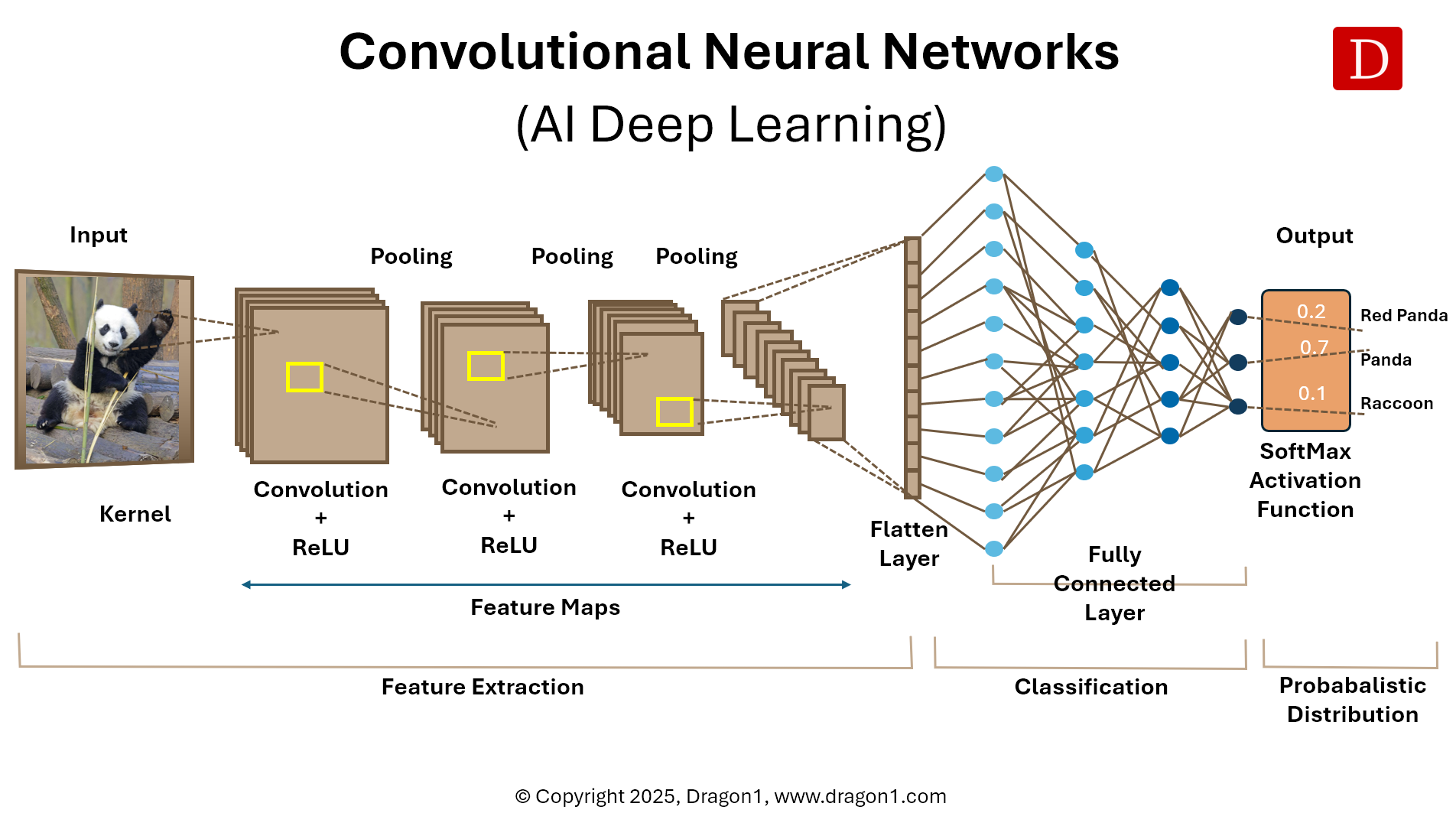
So far, we've focused on the feature extraction part of the CNN. The full architecture can be conceptually broken down into two main stages, as shown in the diagram below.
-
Feature Extraction: This is what we've been discussing. It consists of multiple layers of Convolution (applying filters) and another operation called Pooling. A pooling layer's main job is to shrink the feature maps, making the network more efficient and helping it recognize a feature regardless of its exact position.
-
Classification: After many layers of feature extraction, the network has a rich set of high-level features. The final part of the network, typically called the Fully Connected Layers, acts as the decision-maker. It takes all these abstract features as input and learns to weigh them to produce the final output, like a probability score for "caries: 92%" or "periapical lesion: 78%".
Conclusion
In this lesson, you have conceptually "opened the black box" of how an AI model sees and interprets images. You've moved from understanding an image as a grid of pixels to understanding how a CNN intelligently processes that grid.
Key Takeaways:
- Convolutional Neural Networks (CNNs) are a special type of AI designed for image analysis.
- They use filters to scan an image for simple patterns (like edges and textures) in a process called convolution.
- The output of a convolution is a feature map, which highlights where a specific feature was found in the image.
- CNNs use multiple layers to build a hierarchical understanding: early layers find simple features, and deeper layers combine them to recognize increasingly complex and abstract objects, like anatomical structures or pathologies.
- The entire process involves a feature extraction stage followed by a classification stage to make the final prediction.
Now that you understand how a CNN works conceptually, the next logical question is: how do we create and teach one to perform a specific dental task? Our next lesson, "Outline the process of creating a dental AI model, from data collection and expert annotation to model training," will walk you through that entire workflow.