Hello! Welcome to the next lesson in our module on AI in Radiographic Diagnosis.
In our last lesson, we focused on how to interpret AI outputs for caries detection, looking at visual overlays and confidence scores. We saw how AI answers the question, "Is there something here, and how sure are you?"
Today, we shift our focus from detecting a specific pathology to quantifying a disease state. Our learning outcome is to describe how AI measures alveolar bone loss to assist in classifying periodontal disease severity. We'll explore how AI systems replicate and often enhance the way clinicians assess periodontal health on radiographs, moving from a qualitative assessment to a precise, automated measurement.
The Clinical Benchmark: Radiographic Assessment of Bone Loss
Before we dive into how an AI does it, let's quickly refresh our understanding of the conventional method. As a dental student, you're already familiar with assessing bone levels on radiographs. The goal is to determine the distance between the cementoenamel junction (CEJ) and the alveolar bone crest, and to estimate the percentage of bone loss relative to the total root length.
To revisit these fundamental concepts in a clear, structured way, please watch the following video.
Interpretation of Periodontal Disease Source
This video, from the YouTube channel Radiology 2019, provides an excellent review of how to interpret periodontal disease on radiographs. It will refresh our understanding of the key landmarks and classifications that form the basis for what an AI model is trained to do.
Please watch the sections on the radiographic appearance of the healthy periodontium (02:28 - 04:14) and the classification of bone loss severity (08:21 - 13:21). Pay close attention to how severity is defined by the percentage of bone lost and linked to ADA/AAP classifications.
As the video reinforces, clinical assessment involves:
- Identifying key landmarks: The Cementoenamel Junction (CEJ) and the Alveolar Bone Crest (ABC).
- Establishing a baseline: In health, the ABC is typically 1.5-2.0 mm apical to the CEJ.
- Quantifying loss: Severity is often categorized based on the percentage of bone lost relative to the root length (e.g., <15% for Stage I, 15-33% for Stage II, >33% for Stage III/IV).
This manual process, while standard practice, can be subjective and time-consuming. This is precisely where AI offers a powerful solution.
The AI's Method: From Pixels to Percentages
An AI system automates the measurement of alveolar bone loss by following a logical, multi-step process that mirrors, and in some ways refines, the human approach.
The general workflow is:
- Tooth Segmentation: The AI first identifies and outlines each individual tooth in the radiograph.
- Landmark Localization: It then pinpoints the critical anatomical landmarks for each tooth: the CEJ, the Alveolar Bone Crest (ABC), and the Root Apex (AP).
- Measurement and Calculation: Finally, it measures the distances between these points to calculate the percentage of bone loss and assigns a periodontal stage.
This image from a research paper visually demonstrates the landmark localization step.
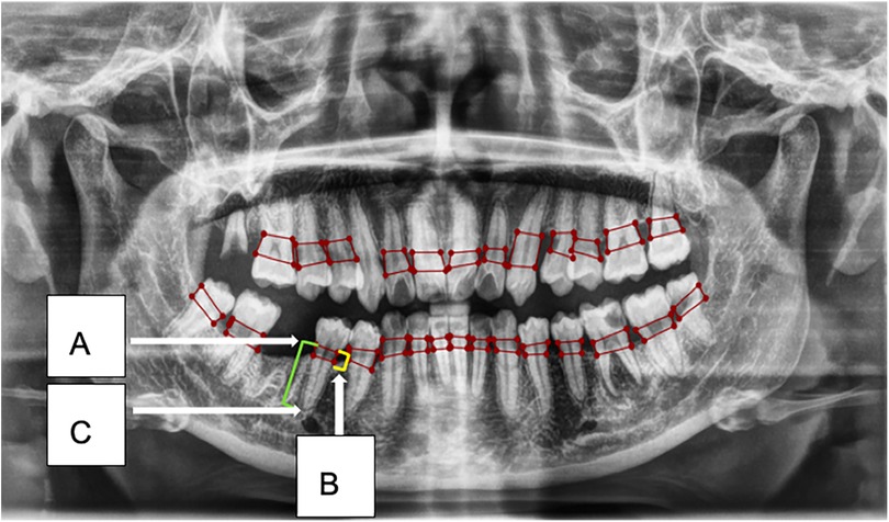
To understand this process more formally, let's look at a recent study that developed an AI model for this exact task.
Artificial intelligence-powered innovations in periodontal diagnosis
This research article, 'Artificial intelligence-powered innovations in periodontal diagnosis,' describes the development of an AI model using YOLOv8 to automate periodontitis staging. It provides a clear, high-level overview of the AI workflow.
Please read section 2.2, focusing on the 'Severity of periodontitis' criteria, which formally defines the staging based on bone loss percentage. Then, read section 2.7, 'AI model development,' especially steps 3 and 4, which explain how the model localizes the CEJ and crest and then uses a formula to calculate the percentage of bone loss for staging.
As the paper describes, the AI is trained to identify the key structures, calculate the percentage of bone loss, and then apply the same staging rules that clinicians use.
A More Advanced Approach: Accounting for Root Curvature
While the method described above is effective, more advanced AI systems have refined the measurement process to improve accuracy. A major challenge in traditional radiographic analysis is that a straight-line measurement from CEJ to apex doesn't account for the natural curvature of a tooth's root. This can lead to inaccuracies in calculating the true percentage of bone loss.
A state-of-the-art AI system called Q-Bone was designed to solve this very problem. It uses an "anatomically-driven" algorithm that measures along the actual curved surface of the root.
Let's examine how it works.
[PDF] an intelligent quantitative system for alveolar bone loss to assist the ...
This paper, 'An intelligent quantitative system for alveolar bone loss...', details the Q-Bone system. We will focus on the specific section that describes its innovative, curvature-based measurement algorithm.
Please read section 2.8, 'Alveolar bone resorption quantification algorithm based on root anatomical curvature.' Focus on the three steps described and the formula for calculating the Alveolar Bone Resorption Ratio (ABRR).
To summarize the brilliant method from the Q-Bone paper:
- Local Coordinate System: For each tooth, the AI establishes an independent coordinate system based on the mesial-distal points of the CEJ. This makes the measurement consistent regardless of tooth angulation.
- Anatomical Correction: It projects the identified alveolar crest point onto the actual segmented outline of the root, ensuring the measurement is taken from the true root surface.
- Curved Measurement: The AI then calculates two lengths along the curved surface of the root:
- Root Length (RL): The curve distance from the CEJ to the root apex.
- Bone Loss (BL): The curve distance from a standardized point (2mm below the CEJ) to the corrected alveolar crest point.
- ABRR Calculation: It uses these values to calculate the Alveolar Bone Resorption Ratio (ABRR) with the formula:
- Staging: Finally, it applies the standard staging criteria to the calculated ABRR percentage:
- Stage I: ABRR ≤ 15%
- Stage II: 15% < ABRR ≤ 33%
- Stage III/IV: ABRR > 33%
This anatomically-aware method provides a more precise and clinically reliable quantification of bone loss than what is feasible with manual, "eyeball" estimation or simple linear tools.
Visualizing the AI's Analysis
Just like with caries detection, the AI presents its findings in a clinically intuitive way. It's not enough to perform the calculation; the system must communicate the results effectively to the clinician.
The Q-Bone system, for instance, includes a comprehensive visualization interface. Let's look at the description in the paper and an example from a commercial product.
[PDF] an intelligent quantitative system for alveolar bone loss to assist the ...
Returning to the Q-Bone paper, let's see how it presents its findings to the user.
Please look at Figure 7 and read the short accompanying text in section 3.5, 'Interactive visualization system for alveolar bone resorption quantification.' Notice the combination of color-coded overlays on the radiograph and the structured table listing the ABRR and stage for each tooth.
This integrated view allows a clinician to instantly see which teeth are affected, the precise level of bone loss calculated by the AI, and the corresponding periodontal stage.
Here is another example from a different AI software:

These user interfaces combine visual overlays with quantitative data, providing you with a comprehensive and efficient "second opinion" to confirm and enhance your own diagnostic assessment. Studies on these systems, such as the ones we've reviewed, consistently show a very high level of agreement between the AI's staging and that of expert periodontists, highlighting their reliability as diagnostic aids.
Conclusion
In this lesson, we have detailed the process by which an AI system measures alveolar bone loss to assist in classifying periodontal disease. We've moved from the foundational clinical principles to the sophisticated, automated algorithms that are transforming this area of diagnostics.
Key Takeaways:
- AI automates the radiographic assessment of periodontal disease by replicating and refining the clinical measurement of bone loss.
- The core process involves tooth segmentation, landmark localization (CEJ, alveolar crest, apex), and calculation of the bone loss percentage.
- Advanced systems use curvature-based algorithms to measure along the true anatomical surface of the root, improving accuracy over simple linear estimates.
- The AI uses the calculated bone loss percentage to automatically assign a periodontal stage based on established clinical guidelines (e.g., AAP/EFP classification).
- The results are presented through intuitive visual interfaces that combine graphical overlays with quantitative data tables, allowing for rapid clinical interpretation.
In our next lesson, we will explore another application of AI in radiographic analysis: describing AI applications in assessing restoration quality and detecting recurrent decay. This will bring us back to a detection task, but one that requires the AI to understand the complex interface between natural tooth structure and restorative materials.