Hello! Welcome to the first lesson in our module on AI in Radiographic Diagnosis.
In our last module, we concluded by discussing how to relate AI performance metrics to clinical utility. We used caries detection as a prime example, framing AI as a powerful screening tool with high sensitivity, excellent at ruling out healthy surfaces and flagging suspicious areas for your expert review.
Today, we'll peel back the layers and look "under the hood." Our goal is to describe how AI detects carious lesions on bitewing and periapical radiographs. We will move from the what (AI as a screening tool) to the how, exploring the computational steps the system takes to go from a standard radiograph to a potential diagnosis.
Why Does Dentistry Need AI for Caries Detection?
As a dental student, you are acutely aware of the challenges in radiographic interpretation. Even with standardized techniques, diagnosis can be subjective. What one clinician sees as an incipient lesion, another may dismiss as an artifact. This variability is a well-known issue in dentistry.
To set the stage, let's watch a brief segment from a TEDx talk that highlights this exact problem and positions AI as a solution.
Eradicating the Dental Divide with AI: new era of preventive care | Florian Hillen | TEDxBoston
In this clip from 'Eradicating the Dental Divide with AI,' Florian Hillen, CEO of VideaHealth, discusses the diagnostic challenges dentists face and how AI can significantly improve accuracy.
Please watch from 02:13 to 04:35 and from 09:00 to 09:40. Pay attention to the discussion on diagnostic variability and the specific claim about how much AI can reduce missed cavities.
As the video points out, studies show that AI can reduce missed caries by a significant margin. But how does it achieve this? The answer lies in a specific type of AI designed for image analysis: the Convolutional Neural Network (CNN).
The AI's "Eye": Convolutional Neural Networks
You'll recall from our second module that AI models process images not as pictures, but as numerical data—a grid of pixels, each with a value representing its shade of grey. A Convolutional Neural Network (CNN) is a deep learning architecture that is exceptionally good at finding patterns within this numerical data.
To understand how CNNs are applied to caries detection, let's read a short excerpt from a review paper on this topic.
[PDF] Transforming Dental Caries Diagnosis Through Artificial Intelligence ...
This article, 'Transforming Dental Caries Diagnosis Through Artificial Intelligence-Based Techniques,' provides an excellent overview. We'll focus on the section explaining the role of CNNs.
Please read the first two paragraphs of the 'Review' section, under the sub-heading 'Convolutional Neural Networks in Image Analysis and Caries Detection.' Focus on how CNNs learn to find features directly from images.
As the text explains, a CNN works by applying a series of filters to an image to learn a "spatial hierarchy of features." Think of it this way:
- The first layers of the network might learn to detect very simple features, like edges or changes in brightness.
- Subsequent layers combine these simple features into more complex ones, like textures or simple shapes.
- Deeper layers learn to recognize even more complex and abstract patterns, such as the characteristic triangular shape of an interproximal lesion or the subtle diffuse radiolucency of an occlusal lesion in dentin.
Through training on thousands of radiographs labeled by expert dentists, the CNN learns which specific combination of features corresponds to a carious lesion, distinguishing it from healthy enamel, dentin, restorations, or image artifacts.
The Automated Detection Workflow
The CNN's pattern-matching ability is the core engine, but it's just one part of a larger, multi-step process. An AI system follows a systematic workflow to analyze a radiograph.
The paper we just looked at provides a fantastic, clear breakdown of this process.
[PDF] Transforming Dental Caries Diagnosis Through Artificial Intelligence ...
Let's return to the same article and read the section that details the entire workflow, from acquiring the image to the final classification.
Please read the entire section titled 'Image processing and analysis techniques for identifying caries lesions.' This section breaks down the process into clear, logical steps.
Based on that reading, let's walk through the workflow. This entire sequence is what happens in the seconds after you upload a radiograph to an AI system.
-
Image Acquisition: The process starts with a digital image, typically a bitewing or periapical radiograph. The quality of this input image is crucial.
-
Pre-processing: The AI first "cleans" the image to make it easier to analyze. This can involve:
- Noise Reduction: Smoothing out graininess or digital artifacts that could be mistaken for pathology.
- Contrast Enhancement: Adjusting the image to maximize the visual difference between healthy tooth structure and demineralized areas, making potential lesions more distinct.
-
Segmentation: This is a critical step. The AI identifies and isolates the teeth from the background (e.g., alveolar bone, soft tissue shadows, empty space). This ensures the subsequent analysis focuses only on the relevant anatomy, preventing a shadow on the bone from being mistaken for a lesion on a tooth.
-
Feature Extraction: Now, the trained CNN gets to work on the segmented teeth. It scans the tooth structures and extracts the key features it has learned to associate with caries, such as:
- Intensity features: The radiolucency (darker pixels) of a demineralized area.
- Texture features: The specific pattern and variation of pixels within the lesion.
- Shape features: The size and geometry of the suspicious area.
-
Classification: Using the features extracted in the previous step, a classifier (the final layers of the neural network) makes a decision. It calculates the probability that the region represents a carious lesion versus healthy tooth structure.
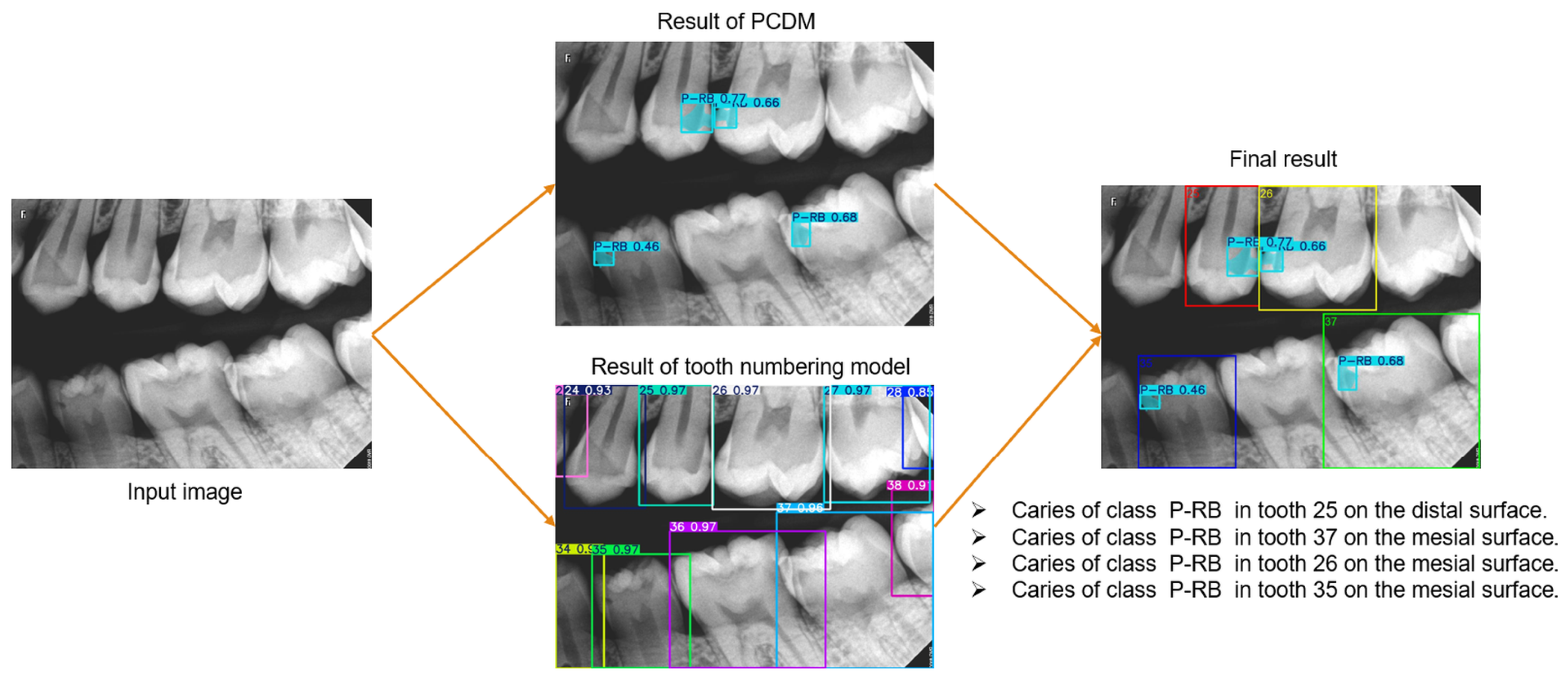
This entire workflow is visualized perfectly in the following image.
Real-World Application
This process isn't just theoretical; it's the foundation for commercial AI software used in dental practices today. Tools like Pearl®, Overjet, and Denti.AI® all use variations of this deep learning workflow to provide real-time analysis of radiographs.
Another research paper provides concrete examples of this process on bitewing and periapical radiographs, demonstrating the high level of performance these models can achieve.
[PDF] Caries detection using deep learning and convolutional neural ...
Let's look at the paper 'Caries detection using deep learning and convolutional neural networks...' for some specific examples.
Please read the short sections titled 'Case Study 1: Convolutional Neural Networks for Bitewing Radiographs' and 'Study 3: U-Net Segmentation and Periapical Radiographs.' Notice the high accuracy and segmentation scores reported.
As these studies show, when applied to bitewings, CNNs can achieve high accuracy in detecting even early-stage lesions. When applied to periapicals, specialized architectures like U-Net can precisely segment (outline) the boundaries of the lesion, providing valuable information about its size and extent.
Conclusion
In this lesson, we have unpacked the process behind AI-powered caries detection. You now have a conceptual model for how these systems analyze bitewing and periapical radiographs.
Key Takeaways:
- AI detects caries by processing radiographs as numerical pixel data, not by "seeing" them in a human sense.
- Convolutional Neural Networks (CNNs) are the core technology used to learn and identify the visual patterns of caries from thousands of expert-labeled examples.
- The detection process is a multi-step workflow:
- Pre-processing to clean the image.
- Segmentation to isolate the teeth.
- Feature Extraction where the CNN finds patterns related to caries.
- Classification to decide if a lesion is present.
- This workflow is the basis for commercial AI tools that assist dentists in real-time radiographic analysis.
Now that you understand how an AI model finds caries, our next lesson will focus on a crucial follow-up question for you as a clinician: How do you read and use the AI's output? We will explore how to interpret AI-generated caries detection outputs, including confidence scores and visual overlays, and integrate them into your diagnostic thinking.