Welcome to the second lesson in our neuroanatomy module. In our previous session, we mapped the functional areas of the cerebral cortex, identifying the primary somatosensory cortex in the parietal lobe as the brain's destination for bodily sensations. This naturally raises the question: how does information about touch, pain, or body position travel from your fingertip all the way to that specific spot in the brain?
Today, we will answer that by charting the course of the nervous system's great sensory superhighways. Our learning outcome is to identify the major ascending tracts—the dorsal column-medial lemniscus and spinothalamic tracts—and their sensory functions. Understanding these pathways is not just an academic exercise; it's the foundation for diagnosing neurological injuries. For your MPT exam and future clinical work, being able to deduce the location of a spinal cord lesion from a patient's specific sensory losses is a critical skill.
The Sensory Nervous System: Two Main Routes to the Brain
Sensory information from the body travels to the brain via two principal, distinct pathways within the spinal cord. Think of them as two types of shipping services: one is an express courier for high-priority, detailed information, and the other is a standard service for more general alerts.
These two pathways are:
- The Dorsal Column-Medial Lemniscus (DCML) Pathway: This is the express courier. It carries information about fine, discriminative touch, vibration, and conscious proprioception (your sense of body position). Its nerve fibers are large and heavily myelinated, ensuring fast and precise signal transmission.
- The Anterolateral System (ALS): This is the standard alert system. Its primary component is the Spinothalamic Tract, which carries protective sensations like pain, temperature, and crude (non-discriminative) touch. Its fibers are smaller and less myelinated, resulting in slightly slower transmission.
The image below provides an excellent side-by-side comparison of these two systems. Notice how they transmit different types of sensations and, crucially, how their paths cross to the opposite side of the brain at different locations.
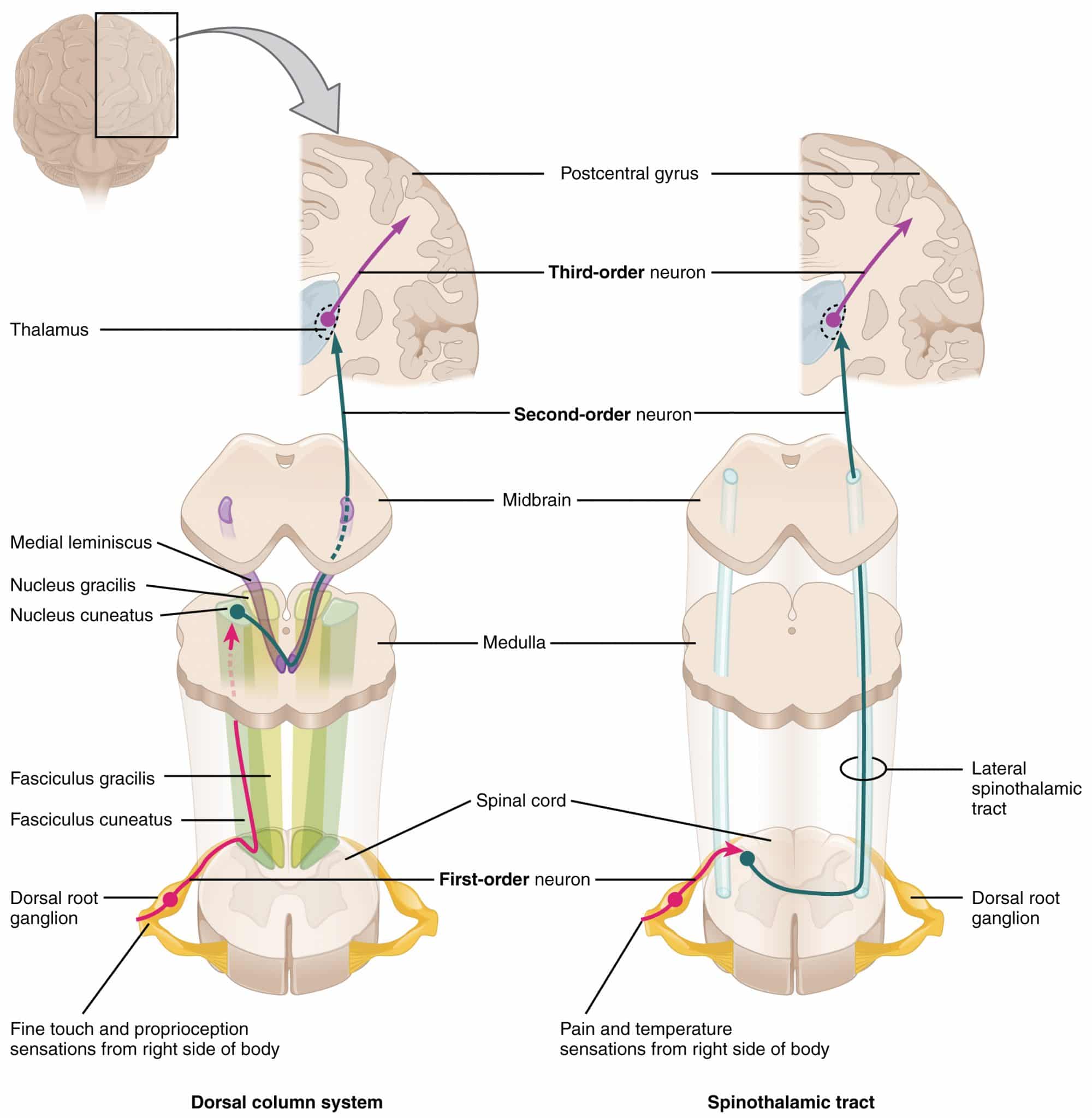
Before we dive into each pathway, it is essential to understand that both systems use a three-neuron relay to get information from the periphery to the cortex:
- First-Order Neuron: Connects the sensory receptor in the skin or muscle to the spinal cord or brainstem. Its cell body is always in the dorsal root ganglion (DRG).
- Second-Order Neuron: Relays the signal from the spinal cord or brainstem to the thalamus. Its axon is the one that decussates.
- Third-Order Neuron: Carries the signal from the thalamus to the primary somatosensory cortex in the postcentral gyrus.
The Express Courier: Dorsal Column-Medial Lemniscus (DCML) Pathway
The DCML pathway is responsible for the sensations that allow us to perform detailed sensory analysis of our environment, like identifying an object in your pocket by feel (stereognosis) or sensing the vibration of a tuning fork.
Sensations Carried:
- Fine (discriminative) touch
- Two-point discrimination
- Conscious proprioception (joint and limb position sense)
- Vibration (pallesthesia)
To trace this pathway in detail, we can consult a clear overview from the StatPearls publishing platform.
Physiology, Sensory System - StatPearls - NCBI Bookshelf
This section from StatPearls provides a concise and accurate description of the DCML pathway, from the peripheral receptors to the cerebral cortex.
Please read the section titled Dorsal Column Pathway. As you read, focus on the three-neuron chain and, most importantly, note where the decussation occurs.
As you just read, the path of the DCML can be summarized as follows:
-
First-Order Neuron: The journey begins at specialized mechanoreceptors (like Meissner's and Pacinian corpuscles). The neuron's axon enters the spinal cord and immediately turns upward, ascending ipsilaterally (on the same side) in the dorsal columns.
- Fibers from the lower body (below T6) travel in the medial Fasciculus Gracilis.
- Fibers from the upper body (T6 and above) travel in the lateral Fasciculus Cuneatus.
-
Second-Order Neuron: The first-order neuron synapses in the medulla, at the Nucleus Gracilis or Nucleus Cuneatus. The axons of the second-order neurons, known as internal arcuate fibers, then decussate (cross over to the contralateral side) in the medulla and ascend through the brainstem as a bundle called the Medial Lemniscus.
-
Third-Order Neuron: The second-order neuron synapses in the Ventral Posterolateral (VPL) nucleus of the thalamus. From there, the third-order neuron projects to the primary somatosensory cortex.
The following animated video provides a fantastic visual walkthrough of this entire pathway.
Spinothalamic Tract and Dorsal Column Medial Lemniscus Pathway | Ascending Tracts of the Spinal Cord
This video from Rhesus Medicine illustrates the journey of sensory information through the DCML pathway.
Please watch the segment from the start of the DCML explanation up to the introduction of the unconscious pathways. Pay close attention to the animated distinction between the Fasciculus Gracilis and Cuneatus, and the point of decussation in the medulla.
The Protective Alert: Anterolateral System (Spinothalamic Tract)
The spinothalamic tract is our fundamental survival pathway. It alerts us to potentially harmful stimuli, mediating the sensations of pain, temperature extremes, and crude touch.
Sensations Carried:
- Pain (nociception)
- Temperature (thermoception)
- Crude (non-discriminative) touch and pressure
Let's return to the StatPearls article to trace this second major pathway.
Physiology, Sensory System - StatPearls - NCBI Bookshelf
This section clearly outlines the path of the spinothalamic tract, highlighting its key differences from the DCML pathway.
Please read the section titled Spinothalamic Pathway. While reading, compare it to the DCML path you just learned. Note the different location of the first synapse and the much earlier point of decussation.
The key difference, as you've noted, is the level of decussation. Let's summarize the spinothalamic path:
-
First-Order Neuron: The journey starts at free nerve endings in the skin. The neuron enters the spinal cord and synapses almost immediately in the dorsal horn (specifically, in a region called the Substantia Gelatinosa).
-
Second-Order Neuron: The axon of the second-order neuron decussates within one or two vertebral levels via the anterior white commissure. It then ascends on the contralateral side of the spinal cord.
- Pain and temperature fibers form the Lateral Spinothalamic Tract.
- Crude touch and pressure fibers form the Anterior Spinothalamic Tract.
-
Third-Order Neuron: The second-order neuron ascends all the way to the VPL nucleus of the thalamus, where it synapses. The third-order neuron then projects to the primary somatosensory cortex.
This next video clip will help you visualize this crucial difference in the pathway.
Spinothalamic Tract and Dorsal Column Medial Lemniscus Pathway | Ascending Tracts of the Spinal Cord
This clip from the same Rhesus Medicine video illustrates the spinothalamic pathway.
Now, watch the section covering the Anterolateral System. Contrast this animation with the DCML pathway you saw earlier. The key visual to grasp is the immediate synapse and crossing over in the spinal cord.

Clinical Application: Decoding Spinal Cord Lesions
Understanding the anatomy of these two tracts is paramount for clinical diagnosis. The different levels of decussation produce a very specific and predictable pattern of sensory loss when the spinal cord is damaged. This is a favorite topic for exam questions.
Let's look at a few classic spinal cord syndromes. The Ninja Nerd video series is renowned for its detailed clinical explanations, which are perfect for reinforcing these concepts for your exam preparation.
- Posterior Cord Syndrome: Caused by damage to the dorsal columns (e.g., from Vitamin B12 deficiency or tertiary syphilis/tabes dorsalis).
- Result: Bilateral loss of proprioception, vibration, and fine touch below the level of the lesion. Pain and temperature sensation are preserved.
- Watch this clip for a clear explanation:
Spinal Cord Lesions: Anterior Cord, Posterior Cord, Central Cord, Brown-Séquard
This clip from Ninja Nerd explains Posterior Cord Syndrome.
Watch the segment on Posterior Cord Syndrome. Note how the lesion's location directly correlates with the loss of DCML functions.
- Central Cord Syndrome: Often caused by a syrinx (a fluid-filled cavity) or trauma that damages the center of the cord.
- Result: The crossing fibers of the spinothalamic tract in the anterior white commissure are damaged first. This causes a bilateral loss of pain and temperature in a "cape-like" distribution across the shoulders and arms. DCML functions are often spared initially.
- This clip explains the mechanism:
Spinal Cord Lesions: Anterior Cord, Posterior Cord, Central Cord, Brown-Séquard
This segment explains the classic "cape-like" sensory loss in Central Cord Syndrome.
Watch from the start of this section to understand how a central lesion specifically impacts the decussating spinothalamic fibers.
- Brown-Séquard Syndrome (Spinal Hemisection): The classic test of your knowledge. Caused by damage to one entire half of the spinal cord (e.g., a stab wound).
- Result (below the lesion):
- Ipsilateral loss of proprioception, vibration, and fine touch (because the DCML tract hasn't crossed yet).
- Contralateral loss of pain and temperature (because the spinothalamic tract has already crossed over in the segments below).
- This is the most complex scenario, and understanding it demonstrates mastery.
- Result (below the lesion):
Spinal Cord Lesions: Anterior Cord, Posterior Cord, Central Cord, Brown-Séquard
This clip breaks down the complex sensory findings in Brown-Séquard Syndrome.
Watch carefully from this section on sensory deficits. The explanation of why pain/temperature loss is contralateral and a few segments below the lesion is crucial.
Conclusion
In this lesson, we have traced the two primary routes for sensory information from the body to the brain. We have established their distinct functions and, most importantly, their unique anatomical paths. The level of decussation is the single most critical feature distinguishing these tracts and is the key to interpreting the clinical signs of spinal cord injury.
Key Takeaways for Exam Revision:
| Feature | Dorsal Column-Medial Lemniscus (DCML) | Spinothalamic Tract (Anterolateral System) |
|---|---|---|
| Primary Function | Fine Touch, Vibration, Conscious Proprioception | Pain, Temperature, Crude Touch |
| Fiber Type | Large, Myelinated (Fast) - Aβ fibers | Small, less myelinated (Slower) - Aδ & C fibers |
| 1st Neuron Synapse | Nucleus Gracilis/Cuneatus (Medulla) | Dorsal Horn (Spinal Cord) |
| Decussation Level | Medulla (as internal arcuate fibers) | Spinal Cord (via anterior white commissure) |
| Lesion Effect | Ipsilateral loss of function below lesion | Contralateral loss of function below lesion |
Now that we understand how sensory signals ascend to the cortex, the next logical step is to examine how motor commands descend from the cortex to produce movement. In our next lesson, we will identify the major descending tracts, such as the corticospinal tract, and explore the clinical effects of their lesions, including the classic signs of upper motor neuron damage.