Welcome back! In our last lesson, we explored the ascending sensory pathways, tracing how information about touch, pain, and body position travels up to the brain. We saw that the location where these tracts cross the midline is crucial for diagnosing spinal cord injuries. Today, we will examine the other side of the coin: how motor commands travel down from the brain to produce movement.
This lesson directly addresses the learning outcome: to identify the major descending tracts (corticospinal, extrapyramidal) and the effects of their lesions. For your MPT exam preparation, a firm grasp of the descending motor systems and the ability to differentiate between upper and lower motor neuron lesions is absolutely essential. These concepts are foundational to nearly all of neurological physiotherapy.
The Motor Command Superhighways
Just as sensory information ascends in organized tracts, motor commands descend from the brain to the spinal cord via a set of pathways known as the descending tracts. The neurons that make up these tracts, originating in the cerebral cortex or brainstem, are collectively called Upper Motor Neurons (UMNs). They are the executive decision-makers. They don't directly contact muscles; instead, they synapse on and control Lower Motor Neurons (LMNs), whose cell bodies are in the spinal cord's anterior horn or in brainstem cranial nerve nuclei. The LMNs are the final common pathway, sending their axons directly out to the muscles.
The descending tracts are broadly divided into two main functional groups:
- Pyramidal Tracts: These originate in the cerebral cortex and are responsible for voluntary, conscious control of muscles. They get their name because they pass through the medullary pyramids of the brainstem.
- Extrapyramidal Tracts: These originate in the brainstem and are responsible for involuntary and automatic control, such as muscle tone, balance, posture, and coordination of head/eye movements.
The diagram below provides a superb high-level overview of this division, which we will explore in detail.

The Pyramidal Tracts: The Architects of Voluntary Movement
The pyramidal system is our direct line of command for conscious action. It consists of two primary tracts: the corticospinal tract for the body and the corticobulbar tract for the head and neck.
To begin, let's get an overview of these two tracts by reading a concise summary.
The Descending Tracts - Pyramidal - TeachMeAnatomy
The "TeachMeAnatomy" article provides a clear, well-structured explanation of the pyramidal tracts. It will introduce you to their origin, course, and primary functions.
In the article, please read the section titled Pyramidal Tracts. Focus on the two subdivisions mentioned, Corticospinal and Corticobulbar, and pay close attention to the general path the neurons take from the cortex downwards.
Corticospinal Tract: Controlling the Body
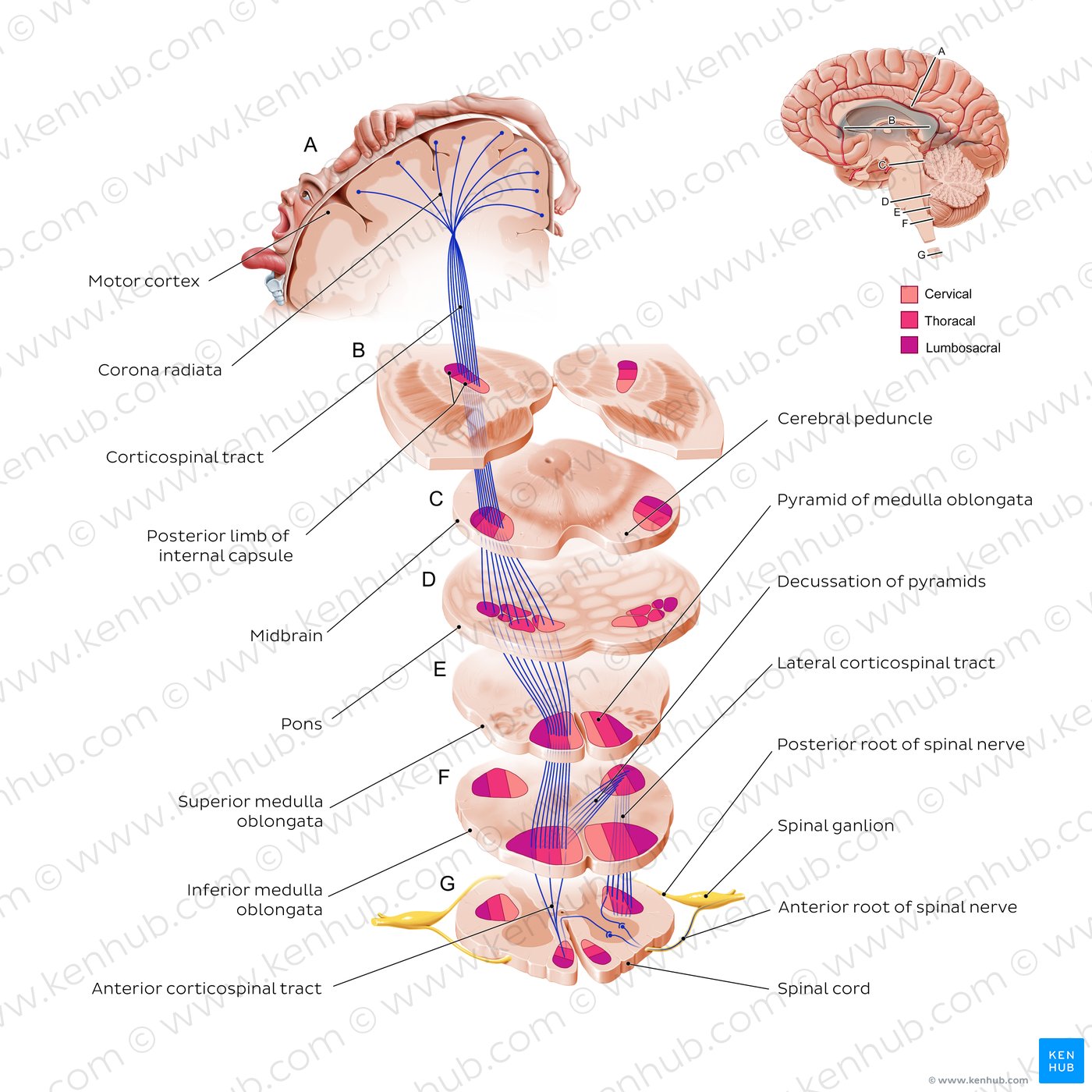
As you just read, the corticospinal tract is the major pathway for voluntary motor control of the trunk and limbs. Let's trace its path, as this is a frequent topic for exam questions.
- Origin: The journey starts in the cerebral cortex, primarily the primary motor cortex, but also the premotor and supplementary motor areas.
- Path: The axons descend through the internal capsule, a dense bundle of white matter. This is a critical point, as it's a common site for strokes ("capsular stroke") which can have devastating motor consequences. The tract then passes through the midbrain (crus cerebri) and pons into the medulla oblongata.
- Decussation: In the lower medulla, at the pyramids, the tract's most important event occurs. About 85-90% of the fibers decussate (cross to the opposite side) to form the Lateral Corticospinal Tract. This tract descends in the lateral part of the spinal cord and controls the contralateral limbs. The remaining 10-15% of fibers do not cross here; they form the Anterior Corticospinal Tract, which descends ipsilaterally to control axial and trunk muscles, with most fibers crossing over at the spinal level they supply.
- Termination: The UMNs of the corticospinal tract synapse on LMNs in the anterior horn of the spinal cord.
This detailed illustration shows the entire course of the corticospinal tract.
Corticobulbar Tract: Controlling the Face, Head, and Neck
The corticobulbar tract follows a similar path from the cortex but terminates on the motor nuclei of the cranial nerves in the brainstem. A key feature is that most cranial nerve nuclei receive bilateral innervation from the corticobulbar tracts (i.e., from both the left and right cerebral hemispheres). This provides a protective redundancy.
However, there are two critical exceptions that are clinically very important:
- Facial Nucleus (CN VII): The part of the nucleus that controls the muscles of the lower face receives only contralateral input.
- Hypoglossal Nucleus (CN XII): This nucleus, controlling tongue movement, also receives primarily contralateral input.
We will see the profound clinical implications of this later in the lesson. This animated video provides a great visual summary of both pyramidal tracts.
Extrapyramidal and Pyramidal Tracts - Descending Tracts of the Spinal Cord | (Includes Lesions)
This clip from Rhesus Medicine provides a clear animation of the pyramidal tracts.
Please watch from the beginning until the start of the extrapyramidal section. Pay close attention to the animated decussation of the corticospinal tract and the explanation of bilateral vs. contralateral innervation in the corticobulbar tract.
The Extrapyramidal Tracts: The Automatic Regulators
While the pyramidal tracts execute our voluntary commands, the extrapyramidal tracts work in the background, originating from brainstem nuclei to automatically regulate posture, balance, muscle tone, and reflexive movements.
Let's read a summary of these four key tracts.
The Descending Tracts of the Central Nervous System | Geeky Medics
This section from "Geeky Medics" provides a quick and effective summary of the four major extrapyramidal tracts, their origins, and their functions.
Please read the entire section under the heading Extrapyramidal tracts. As you read, create a mental map of each tract's name, origin, and primary role.
To summarize the key information for your revision notes:
| Tract | Origin (Brainstem) | Decussation | Primary Function |
|---|---|---|---|
| Rubrospinal | Red Nucleus (Midbrain) | Yes (Contralateral) | Fine motor control, excites upper limb flexors. |
| Reticulospinal | Reticular Formation (Pons & Medulla) | No (Ipsilateral) | Influences voluntary movement and muscle tone. |
| Vestibulospinal | Vestibular Nuclei | No (Ipsilateral) | Controls balance and posture by exciting extensor muscles. |
| Tectospinal | Superior Colliculus (Midbrain) | Yes (Contralateral) | Coordinates head & eye movements to visual stimuli. |
Clinical Correlation: When the Command Center Fails
Understanding the effects of lesions in these tracts is the cornerstone of neurological assessment. Damage to an UMN (the neuron in the descending tract) produces a very different clinical picture than damage to an LMN (the neuron in the spinal cord or cranial nerve nucleus).
The Signs of an Upper Motor Neuron (UMN) Lesion
When the pyramidal tracts are damaged, the inhibitory influence from the cortex is lost. This leads to a characteristic set of "positive" signs due to the disinhibition of lower neuronal circuits.
The cardinal signs of a UMN lesion are:
- Hypertonia: Increased muscle tone, often presenting as velocity-dependent spasticity.
- Hyperreflexia: Exaggerated deep tendon reflexes.
- Clonus: Involuntary, rhythmic muscle contractions in response to stretch.
- Weakness (Spastic Paralysis): The muscle is weak, yet stiff and tight.
- Babinski Sign: Upward extension of the big toe when the sole of the foot is stroked.
This next video from Ninja Nerd is one of the best resources available for understanding why these signs appear. It provides the deep physiological reasoning that is crucial for true mastery beyond simple memorization.
Neurology | Upper Motor Neuron vs Lower Motor Neuron Lesion | UMN vs LMN Lesion
This detailed clip explains the neurophysiological basis of hypertonia and hyperreflexia in UMN lesions.
Watch the segment from the beginning of this section. The key concept to grasp is how the loss of corticospinal input leads to disinhibition of the medullary reticulospinal tract, which in turn causes overactivity in the alpha and gamma motor neurons.
As the video explained, the loss of UMN control unleashes the LMNs and spinal reflexes from their normal cortical inhibition, leading to hypertonia and hyperreflexia. A classic pathological reflex that emerges is the Babinski sign.
Neurology | Upper Motor Neuron vs Lower Motor Neuron Lesion | UMN vs LMN Lesion
This clip explains the mechanism behind the Babinski sign, a hallmark of UMN lesions.
Now, watch this segment explaining the Babinski sign. Understanding why the normal plantar flexion reflex is replaced by dorsiflexion is a high-yield concept for exams.
Contrasting UMN and LMN Lesions
For your MPT exam, you will undoubtedly face questions that require you to differentiate between UMN and LMN lesions. Geeky Medics provides a perfect summary table.
The Descending Tracts of the Central Nervous System | Geeky Medics
This section presents a clear, concise table comparing the clinical signs of UMN and LMN lesions.
Focus on Table 3 and the subsequent paragraph. Note the opposing signs for tone and reflexes and the presence of fasciculations only in LMN lesions.
This final video clip provides a fantastic summary, including the different types of atrophy seen in each lesion type.
Neurology | Upper Motor Neuron vs Lower Motor Neuron Lesion | UMN vs LMN Lesion
This resource explains the difference between disuse and denervation atrophy and provides a final summary of all UMN vs. LMN characteristics.
First, watch the section on atrophy and fasciculations. Then, jump to the final summary chart at the end of the video, which is an excellent tool for rapid revision.
Conclusion
Today, we have charted the descending motor pathways, differentiating between the voluntary pyramidal system and the automatic extrapyramidal system. We traced the critical corticospinal tract from cortex to spinal cord, noting the decussation in the pyramids. Most importantly, we have detailed the profound clinical signs that arise from lesions within these tracts, establishing the classic UMN lesion syndrome and contrasting it with LMN pathology.
Key Takeaways for Exam Revision:
- Pyramidal Tracts (Corticospinal/Corticobulbar): Originate in the cortex for voluntary movement. The corticospinal tract decussates in the medulla.
- Extrapyramidal Tracts (Rubrospinal, etc.): Originate in the brainstem for involuntary control of tone, posture, and balance.
- Lesions of these descending tracts are Upper Motor Neuron (UMN) lesions.
- The comparison below is one of the most important tables in clinical neurology. You must know it thoroughly.
| Physical Sign | UMN Lesion (e.g., Stroke, SCI) | LMN Lesion (e.g., Nerve Root Compression) |
|---|---|---|
| Tone | Hypertonia (Spasticity) | Hypotonia (Flaccidity) |
| Reflexes | Hyperreflexia | Hyporeflexia / Areflexia |
| Atrophy | Mild (Disuse atrophy) | Severe (Denervation atrophy) |
| Fasciculations | Absent | Present |
| Babinski Sign | Present | Absent |
| Paralysis | Spastic | Flaccid |
In our next lesson, we will delve deeper into the control of movement by examining the anatomy of the basal ganglia and the function of its direct and indirect pathways. The basal ganglia are critical structures that modulate the output of the motor cortex, refining our movements and acting as a key component of the extrapyramidal system.