Introduction
Welcome to your first lesson in the Cardiopulmonary Physiotherapy module. As you prepare for your PGET, a strong command of how the cardiovascular and respiratory systems function, both at rest and under stress, is absolutely essential. This foundational knowledge will not only help you answer direct physiological questions but also provide the rationale for many therapeutic interventions you'll be tested on.
In this lesson, we will meet the learning outcome: Describe fundamental cardiovascular and respiratory physiology and their responses to exercise. We'll start by reviewing the core mechanics of the respiratory and cardiovascular systems at rest. Then, we will explore the integrated, dynamic changes that occur in both systems when the body undertakes exercise, a process central to nearly all aspects of physiotherapy.
The Respiratory System at Rest
To understand the body's response to exercise, we must first have a clear picture of the baseline. Your physiotherapy background means you are already familiar with the anatomy of the respiratory system, but a quick review of its physiological function is crucial.
The primary function of the respiratory system is gas exchange: bringing oxygen into the body and removing carbon dioxide. This process involves several coordinated steps: ventilation, diffusion, and transport.
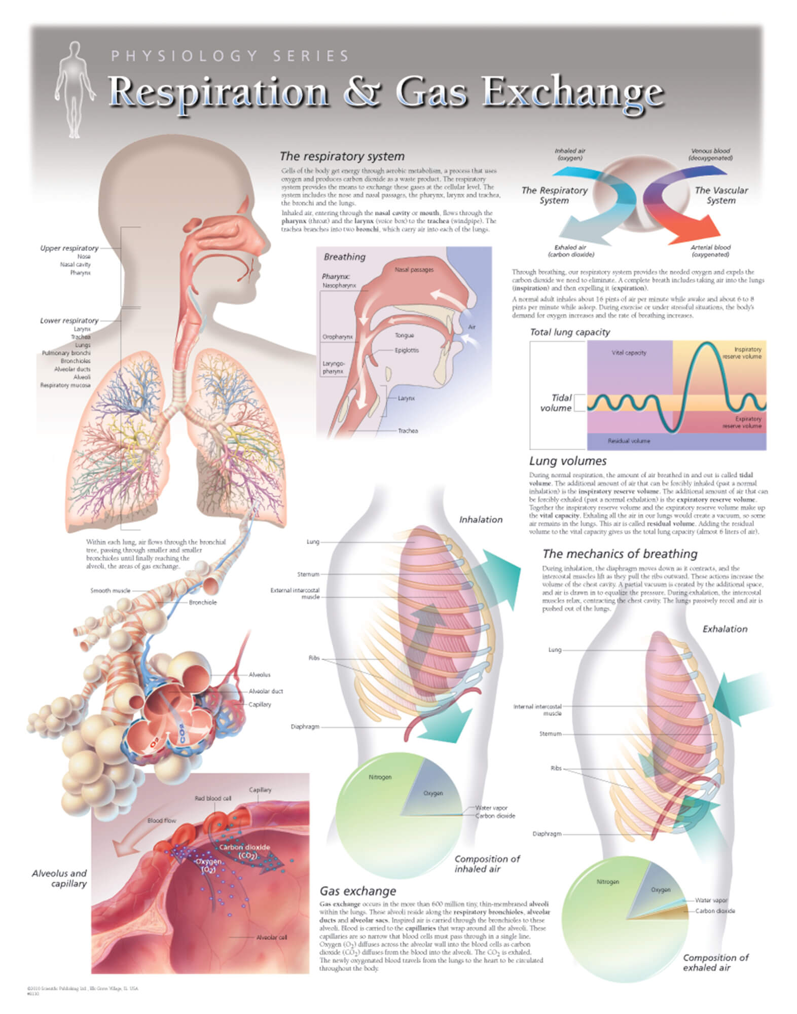
Let's break down the key physiological concepts. The document "Respiratory Physiology" offers a detailed explanation of these fundamentals.
This reading will refresh your understanding of the essential mechanics and principles governing respiration at rest. We will focus on the structures, pressures, volumes, and exchange processes that are critical prerequisites for understanding the response to exercise.
Please read the following sections in the document: Under the ANATOMY heading, review the description of the full respiratory pathway, from the upper airways down to the lungs and pleura, and the dual blood supply. This will re-familiarize you with the key structures. Next, under MECHANISM OF BREATHING, read about the motor pathways and muscles involved in inspiration and expiration. Pay close attention to the roles of the diaphragm and accessory muscles. Under RESPIRATORY PROCESS, study the definitions of key respiratory values like tidal volume, minute volume, and alveolar ventilation. Following that, read the section on Resistance / compliance. Understanding the difference between obstructive and restrictive patterns is a high-yield concept for exams. Finally, focus on the gas exchange process. Read about Diffusion across the alveolar-capillary membrane, the concept of Ventilation/Perfusion (V/Q) matching, and how oxygen is carried in the blood by hemoglobin. Note the factors that shift the oxygen-hemoglobin dissociation curve.
To summarize these resting state concepts:
- Ventilation: Air moves due to pressure gradients created by the contraction and relaxation of respiratory muscles, primarily the diaphragm.
- Compliance & Resistance: The ease of breathing depends on the lung's stretchiness (compliance) and the opposition to airflow (resistance).
- Lung Volumes: At rest, we breathe in a small tidal volume. The volume remaining after a normal exhale is the Functional Residual Capacity (FRC), which acts as an oxygen reserve.
- V/Q Matching: For efficient gas exchange, ventilation (air flow) must be matched with perfusion (blood flow). In the upright lung, both V and Q are greatest at the bases due to gravity, though the matching isn't perfect.
- Oxygen Transport: Most oxygen is transported bound to hemoglobin. The affinity of hemoglobin for oxygen is described by the oxygen-hemoglobin dissociation curve, which can shift based on factors like pH, temperature, and CO₂ levels.
The Cardiovascular System at Rest
The cardiovascular system works in tandem with the respiratory system to deliver oxygenated blood to the tissues and return deoxygenated blood to the lungs. At rest, the heart pumps out a specific volume of blood per minute, known as the cardiac output (CO).
- Heart Rate (HR): The number of times the heart beats per minute, primarily controlled by the autonomic nervous system. At rest, the parasympathetic (vagal) tone dominates, keeping the HR low.
- Stroke Volume (SV): The volume of blood pumped from the left ventricle per beat. It is determined by preload, afterload, and contractility.
- Blood Pressure (BP): The force exerted by circulating blood on the walls of blood vessels. It must be high enough to ensure adequate perfusion of tissues throughout the body.
Integrated Cardiopulmonary Response to Exercise
When exercise begins, the metabolic demand of the working muscles skyrockets. To meet this demand, the respiratory and cardiovascular systems must undergo a series of rapid, coordinated adjustments.
Respiratory Response to Exercise
The most immediate change is an increase in ventilation. This isn't just breathing faster; it's a precisely controlled process to match gas exchange to metabolic need.
This short video provides an excellent summary of the respiratory changes during exercise.
Respiratory Response To Exercise | Respiratory Physiology
Watch this video to get a clear, concise overview of how the respiratory system responds to the increased demands of exercise.
Please watch the entire video (full video). Pay close attention to: The increase in alveolar ventilation. How arterial O_2 and CO_2 levels are maintained. The shift in the oxygen-dissociation curve to the right, facilitating oxygen release to the muscles. How the ventilation-perfusion ratio becomes more uniform across the lung.
In summary, during exercise:
- Increased Alveolar Ventilation: Both the rate and depth of breathing increase, driven by signals from the motor cortex and proprioceptors in moving limbs.
- Stable Arterial Gases: Despite massive increases in consumption and production, the increased ventilation is so well-matched to metabolic rate that arterial and remain remarkably stable during moderate exercise.
- Improved V/Q Matching: Increased cardiac output forces blood into the less-perfused upper zones of the lungs, making the V/Q ratio more uniform and improving the overall efficiency of gas exchange.
- Rightward Shift of O₂-Hb Curve (Bohr Effect): In the exercising muscle tissue, increased temperature, increased , and decreased pH (from lactic acid) cause hemoglobin to release oxygen more readily. This is a critical local adaptation.
Cardiovascular Response to Exercise
The cardiovascular system must dramatically increase oxygen delivery. This is achieved by increasing cardiac output and redistributing blood flow to where it's needed most.
The following video offers a detailed walkthrough of the acute cardiovascular adjustments to exercise. It ties together many concepts you are familiar with, such as the autonomic nervous system, muscle pumps, and the Frank-Starling mechanism.
Acute Cardiovascular Response to Aerobic Exercise
This video explains the key mechanisms the cardiovascular system uses to respond to the onset of aerobic exercise. It provides a strong physiological basis for understanding exercise capacity and limitations.
This is a longer video, so focus on these key segments: Anticipatory Response and ANS: Watch the section explaining the initial changes driven by the autonomic nervous system, including the rise in heart rate, contractility, and systemic vasoconstriction. Venous Return: Pay close attention to the role of the skeletal and respiratory pumps in increasing the amount of blood returning to the heart (venous return). This is a mechanically elegant solution that links movement directly to cardiac filling. Blood Flow Redistribution: Understand how blood flow is shunted. Watch the segment from regulating blood flow which shows blood being directed away from organs like the gut and towards the working muscles and skin. Frank-Starling Mechanism: This is a crucial concept. Watch the explanation of how increased venous return leads to a more forceful contraction, from the Frank-Starling mechanism to the end of the graphical representation. Local Muscle Response: Finally, see how the active muscles themselves influence their own blood supply through metabolic vasodilation and how efficiently they extract oxygen, as described by the arterial-venous oxygen difference.
To solidify these concepts, let's also review them in a text format, which can be helpful for creating your exam notes.
Exercise Physiology - StatPearls - NCBI Bookshelf - NIH
This article from StatPearls provides a concise, system-by-system summary of the physiological responses to exercise.
Please read the following two sections: Cardiovascular System: Read the entire section, starting from this paragraph. It covers the increase in cardiac output, blood flow redistribution, and changes in oxygen transport by red blood cells. Respiratory System: Read the entire section, starting from this paragraph. It reinforces the points from the video about increased ventilation and efficient CO₂ removal.
Synthesis: The Fick Equation
The relationship between the cardiovascular and respiratory systems in delivering oxygen is elegantly summarized by the Fick Equation:
Where:
- is the rate of oxygen consumption by the body's tissues.
- is the cardiac output (), representing central oxygen delivery.
- is the arteriovenous oxygen difference, representing the amount of oxygen extracted by the tissues from the blood (peripheral uptake).
During exercise, the body increases by increasing both (a central cardiovascular adaptation) and the difference (a peripheral adaptation in the muscles). This equation is a powerful tool for understanding overall cardiorespiratory fitness.
Conclusion
In this lesson, we have reviewed the fundamental physiology of the respiratory and cardiovascular systems and detailed their integrated response to the challenge of exercise. Understanding these mechanisms is not just academic; it is the basis for exercise prescription, patient monitoring, and interpreting signs of cardiorespiratory distress.
Key Takeaways:
- Goal of the Response: To increase oxygen delivery and carbon dioxide removal to meet the elevated metabolic demands of exercising muscles.
- Cardiovascular Changes:
- Central Command & ANS: Increased sympathetic drive raises heart rate and myocardial contractility.
- Cardiac Output: Increases dramatically due to rises in both HR and SV.
- Stroke Volume: Enhanced by increased venous return (via muscle/respiratory pumps) and a more forceful contraction (Frank-Starling mechanism).
- Blood Flow Redistribution: Blood is shunted from viscera to active muscles, skin (for cooling), and the heart.
- Respiratory Changes:
- Ventilation: Increases in both rate and depth to match metabolic demand, keeping arterial blood gases stable.
- Gas Exchange Efficiency: V/Q matching improves as the entire lung becomes better perfused.
- Oxygen Unloading: The Bohr effect (rightward shift of the O₂-Hb curve) in active muscles facilitates the release of oxygen from hemoglobin.
In our next lesson, we will build directly on this foundation. We will learn how to perform a cardiopulmonary physiotherapy assessment, which involves techniques like auscultation and interpreting clinical data. Your knowledge of the normal physiological responses we've discussed today will be the critical benchmark against which you'll identify signs of dysfunction.