Welcome back! In our last lesson, you learned a general framework for synthesizing lab results from multiple panels in an undifferentiated patient. We moved from looking at isolated numbers to seeing the connections between them, guided by the patient's clinical story.
Today, we take that skill and apply it to some of the most common and critical situations you'll face in the hospital. The goal is to move beyond general principles and learn to recognize the specific laboratory "fingerprints" of major clinical syndromes. When a patient is critically ill, quickly recognizing these patterns can be life-saving.
Our learning outcome for this lesson is to apply an integrated approach to lab interpretation in common clinical syndromes. We will focus on three high-yield examples:
- Acute Kidney Injury (AKI): Differentiating the cause by synthesizing serum and urine labs.
- Sepsis: Using a constellation of lab findings to identify life-threatening organ dysfunction.
- Acute Liver Failure (ALF): Recognizing the lab triad that defines this medical emergency.
By the end of this lesson, you will be able to spot these syndromes by their characteristic lab patterns, a crucial step toward formulating a rapid management plan.
1. The Signature of Acute Kidney Injury (AKI)
Acute Kidney Injury is an abrupt decline in kidney function that you will encounter constantly in clinical practice. The diagnosis itself is based on lab values, but the real clinical challenge is determining the cause. The approach hinges on classifying the AKI into one of three categories: prerenal, intrinsic renal, or postrenal.
Let's start by watching a detailed overview of this classification.
Acute Kidney Injury (AKI) | Clinical Medicine
This video from Ninja Nerd provides a superb introduction to the definition and classification of AKI. It clearly lays out the pathophysiology behind the three main types, which is the foundation for understanding the lab patterns.
Watch the segment from 00:43 to 20:18. Focus on understanding the core problem in each category: Prerenal: A problem with blood flow to the kidney. Intrinsic: A problem within the kidney itself (tubules, glomeruli, etc.). Postrenal: An obstruction of urine flow after the kidney.
Synthesizing Labs to Determine the Cause of AKI
Once you've diagnosed AKI based on a rise in creatinine or a drop in urine output, the next step is a systematic investigation.
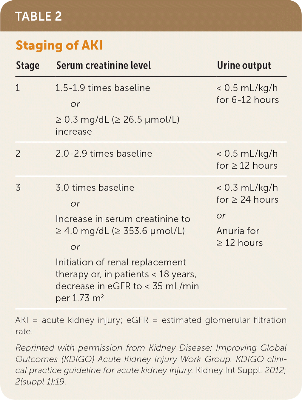
The typical workflow is as follows:
- Rule out postrenal AKI: The first and easiest cause to exclude is obstruction. A simple bladder scan or renal ultrasound can identify a distended bladder or hydronephrosis.
- Differentiate prerenal vs. intrinsic AKI: This is where lab synthesis becomes critical. If there's no obstruction, the cause is either a perfusion problem (prerenal) or damage to the kidney tissue itself (intrinsic).
In prerenal AKI, the kidney is healthy but isn't getting enough blood. It responds appropriately by desperately trying to hold onto salt and water to restore volume. In intrinsic AKI (most commonly Acute Tubular Necrosis, or ATN), the tubules are damaged and can't do their job of reabsorbing salt and water.
This difference in pathophysiology creates distinct laboratory patterns. Let's watch the key segment that explains how to use these patterns for diagnosis.
Acute Kidney Injury (AKI) | Clinical Medicine
This next clip is the most important part of the video for our purposes. It explains exactly how to synthesize serum and urine labs to distinguish between prerenal and intrinsic AKI.
Watch the section on the diagnostic workup from 1:28:21 to 1:40:43. Pay close attention to the rationale behind each test: Fractional Excretion of Sodium (FENa): Is the kidney wasting or conserving sodium? Urine Osmolality: Is the kidney concentrating or diluting urine? BUN:Creatinine Ratio: Why does this ratio go up in prerenal states? Urinalysis Microscopy: What do muddy brown casts signify?
Here is a summary of the key differentiating patterns between the two most common causes of AKI:
| Lab Finding | Prerenal AKI ("Dry") | Intrinsic AKI (ATN - "Damaged") | Rationale |
|---|---|---|---|
| BUN:Cr Ratio | > 20:1 | < 15:1 | In prerenal states, the slow flow in the tubules allows for more urea (BUN) to be reabsorbed along with water, raising the BUN disproportionately to creatinine. |
| Urine Na+ / FENa | Low (< 20 mEq/L) / < 1% | High (> 40 mEq/L) / > 2% | A healthy kidney facing low perfusion (prerenal) avidly reabsorbs sodium. A damaged tubule (ATN) cannot, so it wastes sodium into the urine. |
| Urine Osmolality | High (> 500 mOsm/kg) | Low (< 350 mOsm/kg) | A healthy kidney concentrates urine to save water. Damaged tubules lose this ability, leading to isosthenuria (urine osmolality similar to plasma). |
| Urine Sediment | Bland / Hyaline casts | Muddy brown granular casts | These casts are pathognomonic for ATN and are formed from sloughed, dead tubular epithelial cells. |
Test your understanding!
A 72-year-old man with a history of severe heart failure is admitted for shortness of breath. His baseline creatinine is 1.2 mg/dL. On admission, his creatinine is 2.4 mg/dL and his BUN is 60 mg/dL. His urine sodium is 10 mEq/L and his urine osmolality is 550 mOsm/kg.
Based on these labs, what is the most likely type and cause of his AKI?
Show answer
This is a classic picture of prerenal AKI due to cardiorenal syndrome.
- BUN:Cr ratio is 60:2.4 = 25:1 (> 20:1).
- Urine sodium is low (< 20 mEq/L), indicating avid sodium retention.
- Urine osmolality is high (> 500 mOsm/kg), indicating the kidneys are appropriately concentrating urine.
The labs show a healthy kidney that is responding to a state of poor perfusion caused by the patient's failing heart.
2. The Pattern of Sepsis and Septic Shock
Sepsis is not defined by a single lab test but by a clinical syndrome: life-threatening organ dysfunction caused by a dysregulated host response to infection. Your job as an interpreter of labs is to recognize the signs of that organ dysfunction.
Sepsis and Septic Shock - Critical Care Medicine
The Merck Manual provides a concise and clinically relevant overview of sepsis. We will use it to understand the diagnostic criteria and the constellation of lab findings.
First, read the 'Diagnosis' section to understand the clinical context and scoring systems like qSOFA and SOFA. Then, read the section immediately following the SOFA score table that begins 'CBC, arterial blood gases (ABGs)...'. This part details the specific lab abnormalities that signify organ dysfunction in sepsis.
The diagnosis of sepsis starts with a suspicion of infection plus evidence of organ failure. The Sequential Organ Failure Assessment (SOFA) score formalizes this by assigning points for dysfunction across various organ systems, many of which are defined by lab values.
| Table: Sequential Organ Failure Assessment (SOFA) Score |
|---|
 |
| The SOFA score quantifies organ dysfunction across respiratory, coagulation, liver, cardiovascular, CNS, and renal systems. A rising score indicates worsening sepsis. |
Here is the classic laboratory "fingerprint" of a patient developing sepsis, with each finding representing a piece of the organ dysfunction puzzle:
- Inflammatory Response (CBC):
- Leukocytosis (>12,000) with a "left shift" (increased immature neutrophils/bands) is the classic response.
- Leukopenia (<4,000) is also possible and is a more ominous sign, suggesting the bone marrow is overwhelmed.
- Coagulation (CBC & Coags):
- Thrombocytopenia (platelets < 100,000) is very common due to platelet consumption in microthrombi. Worsening thrombocytopenia is a poor prognostic sign.
- Tissue Perfusion (Metabolic):
- Elevated Lactate (>2 mmol/L): This is a critical marker of tissue hypoperfusion and anaerobic metabolism. A persistently high lactate defines septic shock.
- Acid-Base (ABG/VBG):
- Early on, you may see respiratory alkalosis as the patient hyperventilates (tachypnea).
- As shock progresses, this gives way to a high anion gap metabolic acidosis (HAGMA) due to the rising lactate.
- Kidney Function (BMP):
- Rising Creatinine and BUN: This is the AKI component of sepsis, a very common complication. The pattern can be prerenal initially (due to hypotension) but often progresses to intrinsic ATN ("septic ATN").
- Liver Function (LFTs):
- Elevated Transaminases (AST/ALT) and Bilirubin: This reflects "shock liver" or ischemic hepatitis from poor perfusion. It's typically a mild to moderate elevation.
No single one of these findings is sepsis. It is the combination of these abnormalities in a patient with a suspected infection that constitutes the syndrome.
3. The Triad of Acute Liver Failure (ALF)
Acute Liver Failure is a rare but devastating syndrome. Unlike chronic liver disease, it occurs in patients without pre-existing cirrhosis. The diagnosis is made by a specific and dramatic triad of laboratory and clinical findings.
Chalk Talk: Acute Liver Failure
This 'Chalk Talk' video from The ICU Curriculum is a concise, high-yield summary of ALF. It clearly presents the diagnostic criteria and the key management principles, which are all tied to lab monitoring.
First, watch the definition of ALF from 00:25 to 02:25. Memorize the three components of the diagnostic triad. Then, watch the 'Management' section from 06:05 to 10:58. Focus on the table that links liver function, what happens in failure, how it's monitored with labs, and the clinical intervention.
As the video explains, ALF is defined by three components:
- Evidence of Acute Liver Injury: This is shown by massively elevated aminotransferases (AST/ALT), often in the thousands.
- Impaired Synthetic Function (Coagulopathy): The liver can no longer produce clotting factors. This is the key diagnostic marker, defined as an INR ≥ 1.5.
- Hepatic Encephalopathy: The liver can no longer clear toxins like ammonia, which build up and affect the brain, causing confusion, lethargy, and asterixis (a flapping tremor).
This image provides a great visual summary of how the failure of multiple liver functions leads to the clinical and laboratory syndrome we see.

When managing a patient with ALF, you are essentially monitoring the failure of each of the liver's core functions through lab tests:
- Synthetic Function: Monitor INR and albumin. The INR reflects acute changes; albumin reflects more chronic function.
- Detoxification: Monitor ammonia levels, which correlate with the severity of encephalopathy.
- Metabolic Regulation: Monitor glucose frequently. A failing liver can't perform gluconeogenesis, leading to profound hypoglycemia.
- Excretion: Monitor bilirubin. Jaundice is a prominent physical sign.
Test your understanding!
A 19-year-old is brought to the ED after a suicide attempt with a massive acetaminophen overdose. She is lethargic and confused. Her labs are: AST 8,000, ALT 6,500, INR 2.5, Glucose 45 mg/dL.
- Does she meet the criteria for Acute Liver Failure?
- Which lab result best explains her confusion?
- What other lab test would you immediately order to quantify this?
Show answer
- Yes. She has evidence of acute liver injury (massive AST/ALT), coagulopathy (INR > 1.5), and encephalopathy (confusion).
- Her confusion is due to hepatic encephalopathy.
- You would immediately order a serum ammonia level.
Conclusion
Today, you've learned to recognize the laboratory "fingerprints" of three critical clinical syndromes. This skill goes beyond just identifying abnormal values; it's about seeing the integrated pattern that tells a story of organ failure.
Key Takeaways:
- AKI Pattern: The workup hinges on differentiating prerenal (high BUN/Cr, low FENa, concentrated urine) from intrinsic ATN (low BUN/Cr, high FENa, dilute urine, muddy brown casts).
- Sepsis Pattern: This is a constellation of findings reflecting multi-organ dysfunction in response to infection. Look for inflammatory markers (WBC), cell-line depression (platelets), metabolic derangement (lactate, acidosis), and signs of kidney and liver injury.
- ALF Pattern: This is a specific triad: evidence of severe hepatocellular injury (high AST/ALT), impaired synthesis (INR ≥ 1.5), and hepatic encephalopathy.
Preview of the Next Lesson:
Now that you can recognize these patterns and make a presumptive diagnosis, the next logical question is: "What do I do about it?" In our next lesson, we will focus on formulating a diagnostic or monitoring plan based on initial or evolving laboratory abnormalities. You'll learn how to use serial lab testing to track a patient's response to treatment and anticipate complications.