Hello! Welcome back to our module on acid-base balance.
In our last lesson, you learned to identify the four core components of an Arterial Blood Gas (ABG) report—pH, PaCO₂, PaO₂, and HCO₃⁻—and their normal ranges. This is the essential first step of ABG interpretation.
Today, we will take the crucial next step. Using those values, you will learn to determine the primary acid-base disturbance. This means answering the core question: "Is the problem respiratory or metabolic, and is it an acidosis or an alkalosis?" Mastering this systematic approach will allow you to quickly identify the root cause of a patient's acid-base imbalance, a fundamental skill in clinical medicine.
A Systematic Approach to Finding the Primary Disturbance
The key to interpreting ABGs without getting overwhelmed is to follow a consistent, step-by-step process. For identifying the primary disturbance, we will use a three-step method.
The article "Arterial blood gases made easy" outlines this logic clearly.
Arterial blood gases made easy - PMC
This article from the National Center for Biotechnology Information (NCBI) provides a concise, logical framework for ABG interpretation. We will use its initial steps as the foundation of our method.
Please read the sections "First – look at the pH" and "Second – look at the PaCO2". Notice how this approach quickly narrows down the possibilities by looking at just two values.
Let's expand on that logic and formalize our three-step approach.
Step 1: Look at the pH. Is it Acidosis or Alkalosis?
This is your anchor. The pH tells you the net result of all the buffers and derangements in the blood.
- pH < 7.35 → Acidosis (The patient has acidemia)
- pH > 7.45 → Alkalosis (The patient has alkalemia)
Step 2: Look at the PaCO₂. Does it Explain the pH?
Recall that CO₂ is an acid. The lungs control PaCO₂ by ventilation.
- If the patient has Acidosis (low pH), a high PaCO₂ would be the cause. This is Respiratory Acidosis.
- If the patient has Alkalosis (high pH), a low PaCO₂ would be the cause. This is Respiratory Alkalosis.
If the PaCO₂ change doesn't fit the pH change (e.g., acidosis with a low PaCO₂), the problem isn't respiratory. Let's move to step 3.
Step 3: Look at the HCO₃⁻. Does it Explain the pH?
Recall that HCO₃⁻ is a base. The kidneys control HCO₃⁻ levels.
- If the patient has Acidosis (low pH), a low HCO₃⁻ would be the cause. This is Metabolic Acidosis.
- If the patient has Alkalosis (high pH), a high HCO₃⁻ would be the cause. This is Metabolic Alkalosis.
The Geeky Medics video below provides a great walkthrough of this logic.
ABG Interpretation | Understanding Arterial Blood Gas Analysis - OSCE Guide | UKMLA | CPSA | PLAB 2
This video from Geeky Medics walks through the roles of pH, PaCO₂, and HCO₃⁻ in determining the primary disturbance.
Please watch from 02:11 to 07:39. Pay close attention to how the narrator links a high or low PaCO₂ and HCO₃⁻ back to the pH to determine the cause of the acidosis or alkalosis.
Pattern Recognition: The ROME Mnemonic
For rapid pattern recognition, which aligns with your goal of practical application, a very helpful mnemonic is ROME:
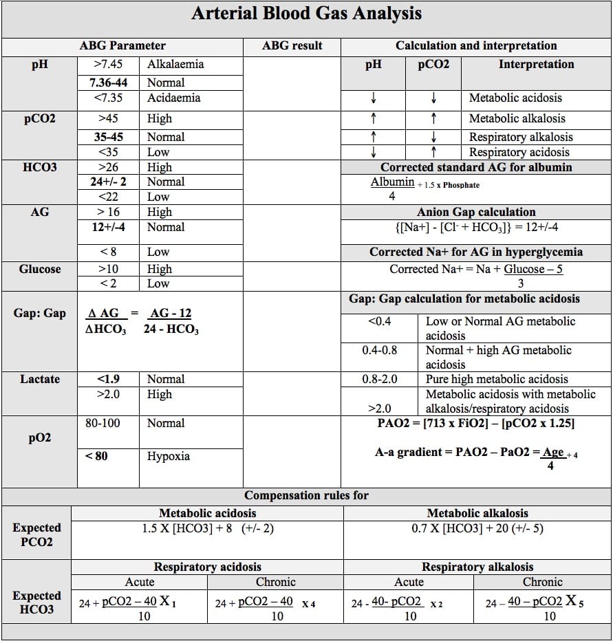
- Respiratory Opposite: In respiratory disorders, the pH and PaCO₂ move in opposite directions.
- Respiratory Acidosis: pH ↓, PaCO₂ ↑
- Respiratory Alkalosis: pH ↑, PaCO₂ ↓
- Metabolic Equal: In metabolic disorders, the pH and HCO₃⁻ move in the same (equal) direction.
- Metabolic Acidosis: pH ↓, HCO₃⁻ ↓
- Metabolic Alkalosis: pH ↑, HCO₃⁻ ↑
This simple rule allows you to glance at an ABG and immediately form a hypothesis.
This chart provides a fantastic visual summary of these patterns and is a great reference to keep handy.
Let's Practice with Clinical Cases
The best way to solidify this is to apply it. We'll use the case studies from Geeky Medics. For each one, use our 3-step method to determine the primary disturbance.
Case 1: Asthma Attack
A 24-year-old asthmatic patient presents with wheezing and shortness of breath. She is breathing very rapidly.
ABG Results:
- pH: 7.49
- PaCO₂: 31 mmHg (Normal: 35-45) Note: original article uses kPa, converted for consistency
- HCO₃⁻: 24 mEq/L (Normal: 22-26)
Test your understanding: Interpret this ABG.
- Step 1 (pH): The pH is 7.49, which is high. This is an alkalosis.
- Step 2 (PaCO₂): The PaCO₂ is 31 mmHg, which is low. A low level of acid would cause alkalosis. This matches the pH.
- Step 3 (HCO₃⁻): The HCO₃⁻ is 24 mEq/L, which is normal. This is not contributing to the problem.
Conclusion: The low PaCO₂ explains the high pH. This is a Respiratory Alkalosis.
Check with ROME: pH is up, PaCO₂ is down. They are moving in opposite directions, confirming a respiratory disorder. This makes clinical sense—her rapid breathing (hyperventilation) from the asthma attack is causing her to "blow off" too much CO₂.
(This corresponds to Case study 2 in the Geeky Medics resource LINK.)
Case 2: Profuse Vomiting
A 72-year-old woman presents with several days of profuse vomiting.
VBG Results (Venous Blood Gas):
- pH: 7.48
- PvCO₂: 50 mmHg (Normal Venous: ~41-51) Note: a VBG is used here; pH and HCO₃⁻ are still reliable for acid-base status.
- HCO₃⁻: 33 mEq/L (Normal: 22-26)
Test your understanding: Interpret this ABG.
- Step 1 (pH): The pH is 7.48, which is high. This is an alkalosis.
- Step 2 (PvCO₂): The PvCO₂ is high. A high level of acid would cause acidosis, not alkalosis. This does not match the pH. (In fact, the high CO₂ suggests the respiratory system might be trying to compensate for the alkalosis by retaining acid).
- Step 3 (HCO₃⁻): The HCO₃⁻ is 33 mEq/L, which is high. A high level of base would cause alkalosis. This matches the pH.
Conclusion: The high HCO₃⁻ explains the high pH. This is a Metabolic Alkalosis.
Check with ROME: pH is up, HCO₃⁻ is up. They are moving in the same (equal) direction, confirming a metabolic disorder. This makes clinical sense—vomiting causes a large loss of stomach acid (HCl), leading to a net increase of bicarbonate in the blood.
(This corresponds to Case study 5 in the Geeky Medics resource LINK.)
Case 3: Diabetic Ketoacidosis (DKA)
A 21-year-old woman presents with vomiting, lethargy, and confusion. Her breathing is deep and rapid (Kussmaul breathing).
ABG Results:
- pH: 7.30
- PaCO₂: 31 mmHg (Normal: 35-45)
- HCO₃⁻: 13 mEq/L (Normal: 22-26)
- Glucose: 486 mg/dL (very high)
Test your understanding: Interpret this ABG.
- Step 1 (pH): The pH is 7.30, which is low. This is an acidosis.
- Step 2 (PaCO₂): The PaCO₂ is 31 mmHg, which is low. A low level of acid would cause alkalosis, not acidosis. This does not match the pH. (Her rapid breathing is an attempt to compensate by blowing off acid).
- Step 3 (HCO₃⁻): The HCO₃⁻ is 13 mEq/L, which is low. A low level of base (or consumption of base by an acid) would cause acidosis. This matches the pH.
Conclusion: The low HCO₃⁻ explains the low pH. This is a Metabolic Acidosis.
Check with ROME: pH is down, HCO₃⁻ is down. They are moving in the same (equal) direction, confirming a metabolic disorder. This is the classic pattern for DKA, where ketoacids consume the body's bicarbonate stores, driving the pH down.
(This corresponds to Case study 1 in the Geeky Medics resource LINK.)
A Brief Preview of Compensation
In the last example (DKA), you may have noticed that the PaCO₂ was also abnormal, even though it wasn't the primary problem. It was low, which would normally cause an alkalosis. Why was it low in a patient with acidosis?
This is compensation. The body's respiratory system detected the metabolic acidosis and tried to fight back by increasing the respiratory rate to "blow off" CO₂, an acid. This is an attempt to raise the pH back towards normal. The respiratory system responds within minutes, whereas the kidneys take hours to days to compensate for a respiratory problem.
We won't go into the details of calculating compensation today, but recognizing that the value not causing the primary problem is often moving in the opposite direction is the first step to understanding this crucial concept.
Conclusion
You have now built upon your knowledge of ABG components to diagnose the four primary acid-base disturbances. This is a high-yield skill that is critical for managing acutely ill patients.
Key Takeaways:
- A 3-step method (check pH, then PaCO₂, then HCO₃⁻) provides a reliable way to find the primary disturbance.
- The ROME mnemonic is a powerful tool for rapid pattern recognition: Respiratory Opposite, Metabolic Equal.
- The four primary simple disturbances are:
- Respiratory Acidosis: Low pH, High PaCO₂
- Respiratory Alkalosis: High pH, Low PaCO₂
- Metabolic Acidosis: Low pH, Low HCO₃⁻
- Metabolic Alkalosis: High pH, High HCO₃⁻
In our next lesson, we will dive deeper into compensation. You will learn to assess whether the body's response to an acid-base disturbance is appropriate and use formulas, such as Winter's formula for metabolic acidosis, to determine if a second, co-existing disorder is present.