Hello! Welcome to your next lesson on renal function.
In our previous session, we established a strong foundation by interpreting the key serum markers of kidney function: BUN, creatinine, and the eGFR. You learned how to use the BUN/creatinine ratio to differentiate between prerenal and intrinsic kidney injury. Now, we'll shift our focus from the blood to the urine itself, adding another essential layer to your diagnostic toolkit.
Today's learning outcome is to interpret urine dipstick results for protein, blood, glucose, ketones, and nitrites. The urine dipstick is a rapid, inexpensive, and powerful screening test performed on millions of patients daily. Mastering its interpretation is a fundamental skill for any clinician, allowing you to quickly spot clues for conditions ranging from urinary tract infections to diabetes and serious kidney disease.
The Urine Dipstick: A First Look
Before we dive into each component, let's get a feel for the test itself. A urine dipstick is a plastic strip with small chemical pads that change color when they react with substances in the urine. The color change is then compared to a reference chart to get a semi-quantitative result.
Interpretation of the Urinalysis (Part 2) - The Dipstick
To see how this works in practice, watch this brief introduction from the 'Interpretation of the Urinalysis (Part 2) - The Dipstick' video by Strong Medicine.
Watch from 0:23 to 2:04. This clip will show you what a dipstick is, how it's used, and how the results are read by comparing the pads to a color chart.
This is a typical reference chart found on the bottle of dipstick strips. You'll become very familiar with these.
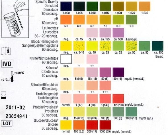
Now, let's break down the most clinically important components one by one.
Glucose and Ketones: Markers of Metabolic State
These two are often interpreted together, as they provide a rapid screen for derangements in glucose metabolism, most notably diabetes mellitus.
Glucose (Glucosuria)
Normally, the kidneys filter glucose at the glomerulus but completely reabsorb it in the proximal tubule.
- Normal Result: Negative.
- Mechanism: The proximal tubule has a maximum capacity for reabsorbing glucose. When the blood glucose level exceeds the renal threshold (typically around 180-200 mg/dL), these transporters are overwhelmed, and glucose "spills" into the urine.
- Clinical Significance:
- Diabetes Mellitus: By far the most common cause. Glucosuria in a patient who isn't known to have diabetes warrants an immediate blood glucose check.
- SGLT-2 Inhibitors: This class of diabetes medication (e.g., empagliflozin, canagliflozin) works by intentionally blocking glucose reabsorption, leading to therapeutic glucosuria.
- Fanconi Syndrome: A rare proximal tubule dysfunction where glucose and other substances are wasted in the urine despite normal blood levels.
Ketones (Ketonuria)
Ketones are produced when the body metabolizes fat for energy instead of carbohydrates.
- Normal Result: Negative.
- Mechanism: Ketones are the byproducts of fatty acid breakdown. The main three are acetoacetate, beta-hydroxybutyrate, and acetone.
- Clinical Significance:
- Diabetic Ketoacidosis (DKA): This is the most urgent diagnosis to consider. A patient with uncontrolled type 1 diabetes can present with extremely high blood sugar, and their dipstick will be strongly positive for both glucose and ketones.
- Other causes: Starvation, prolonged vomiting, extreme exercise, alcoholic ketoacidosis, and very strict ketogenic diets can also produce ketones.
Interpretation of the Urinalysis (Part 2) - The Dipstick
The Strong Medicine video provides excellent clinical context for glucosuria and ketonuria, including a critical limitation of the dipstick test for ketones.
First, watch from 6:59 to 7:57 for a review of glucosuria. Then, watch from 11:44 to 13:20 for the discussion on ketones. Pay close attention to the part about which ketone body the dipstick fails to detect effectively in DKA.
As the video highlighted, the dipstick primarily detects acetoacetate. In DKA, the predominant ketone is beta-hydroxybutyrate. This means that in severe DKA, the dipstick can underestimate the degree of ketosis. It's a crucial clinical pearl.
Test your understanding!
A 22-year-old patient with type 1 diabetes presents to the emergency department feeling unwell, with nausea and abdominal pain. A urine dipstick shows 4+ glucose and 3+ ketones. What is the most likely diagnosis, and what should be your immediate next step?
Show answer
The combination of large amounts of glucose and ketones in the urine is the classic pattern for Diabetic Ketoacidosis (DKA), a medical emergency. Your immediate next steps should be to check a fingerstick blood glucose, obtain serum electrolytes and a blood gas analysis to assess for acidosis, and start treatment with IV fluids and insulin.
Protein: A Sign of Glomerular Trouble
A healthy glomerulus acts as a barrier, preventing large molecules like albumin from passing into the urine. The presence of significant protein is therefore a major red flag for kidney disease.
- Normal Result: Negative or Trace.
- Mechanism: The dipstick is most sensitive to albumin. It is relatively insensitive to other types of proteins, such as the immunoglobulin light chains (Bence Jones proteins) seen in multiple myeloma.
- Clinical Significance:
- A dipstick result of 1+ or greater is considered clinically significant proteinuria and suggests glomerular injury.
- The result is semi-quantitative (Trace, 1+, 2+, 3+, 4+), with higher values indicating more severe protein loss.
- Persistent proteinuria is the hallmark of Chronic Kidney Disease (CKD), often from causes like diabetic nephropathy or hypertension.
- Proteinuria can also be transient, caused by fever, strenuous exercise, or even just standing upright for long periods (orthostatic proteinuria).
It is vital to remember that the dipstick is a screening test for proteinuria. A positive result must be followed up with a quantitative test, such as a urine albumin-to-creatinine ratio (UACR), to accurately measure the amount of protein being lost per day.
Office-Based Urinalysis: A Comprehensive Review
For a practical guide to interpreting proteinuria and a list of potential causes, please review the 'Office-Based Urinalysis' article.
Read the section titled 'PROTEIN'. Pay attention to the table that gives the approximate proteinuria equivalent for each dipstick finding (Trace to 4+). Skim the subsequent table that lists the differential diagnosis for proteinuria to appreciate the breadth of possible causes.
Blood: Hematuria, Hemoglobinuria, or Myoglobinuria?
This is one of the most common abnormal findings and requires careful interpretation.
- Normal Result: Negative.
- Mechanism: The dipstick test detects the peroxidase activity of heme. Heme is the iron-containing component of both hemoglobin (in red blood cells) and myoglobin (in muscle cells).
- Clinical Significance: A positive "blood" result on a dipstick can mean one of three things:
- Hematuria: There are actual red blood cells (RBCs) in the urine. This can be caused by anything from a simple UTI or kidney stone to glomerular disease or bladder cancer.
- Hemoglobinuria: There is free hemoglobin in the urine, but no RBCs. This occurs when RBCs are destroyed within the bloodstream (intravascular hemolysis).
- Myoglobinuria: There is myoglobin in the urine. This occurs when there is significant muscle breakdown (rhabdomyolysis), often from trauma, extreme exertion, or certain drugs.
Because the dipstick can't tell these three apart, a positive dipstick for blood MUST be followed by urine microscopy. If the microscope shows RBCs, you have confirmed hematuria. If the dipstick is positive but the microscope shows no RBCs, you must consider hemoglobinuria or myoglobinuria.
Nitrites and Leukocyte Esterase: The UTI Duo
These two tests are interpreted together as the primary dipstick screen for a Urinary Tract Infection (UTI).
- Normal Result: Both negative.
Leukocyte Esterase (LE)
- Mechanism: LE is an enzyme released by neutrophils (a type of white blood cell).
- Significance: A positive result indicates pyuria (WBCs in the urine), which is a sign of inflammation. While most often caused by a UTI, it can also be seen in non-infectious inflammatory conditions.
Nitrites
- Mechanism: Many common urinary pathogens (especially Gram-negative bacteria like E. coli) contain an enzyme that converts the nitrates normally present in urine into nitrites.
- Significance: A positive nitrite test is highly specific for the presence of these bacteria.
Interpretation as a Pair:
- LE positive + Nitrite positive: High predictive value for a UTI.
- LE positive + Nitrite negative: Suggests a UTI caused by a non-nitrate-reducing organism (e.g., Enterococcus, Staphylococcus) or sterile pyuria (inflammation without infection).
- LE negative + Nitrite positive: Uncommon, but can occur in very early infection before a significant inflammatory response has mounted.
- False-Negative Nitrites: This can happen if the urine hasn't been in the bladder long enough for bacteria to convert nitrates (requires ~4 hours) or if the infecting bacteria lacks the necessary enzyme.
Interpretation of the Urinalysis (Part 2) - The Dipstick
The Strong Medicine video provides a fantastic, clinically nuanced explanation of how to interpret leukocyte esterase and nitrites together.
Watch from 9:45 to 11:44. Focus on why these two tests are considered together and the general guide for interpreting the different combinations of results.
Test your understanding!
A 68-year-old female complains of a 2-day history of painful urination and urinary frequency. Her urine dipstick is positive for leukocyte esterase but negative for nitrites. Does this rule out a UTI? What are some possible explanations?
Show answer
No, this does not rule out a UTI. While a positive nitrite test is very specific, a negative result is common. Possible explanations include:
- UTI with a non-nitrate-reducing organism: Common culprits include Enterococcus faecalis or Staphylococcus saprophyticus.
- Insufficient bladder incubation time: If the patient is urinating very frequently, the bacteria may not have the 4+ hours needed to produce nitrites.
- Sterile pyuria: The inflammation (positive LE) could be due to a non-infectious cause like interstitial cystitis, a kidney stone, or inflammation adjacent to the urinary tract, though UTI is more likely given her symptoms.
A Note on False Positives and Negatives
As you've seen, the dipstick is a screening tool with limitations. Various factors can interfere with the results.
Office-Based Urinalysis: A Comprehensive Review
The AAFP article 'Office-Based Urinalysis' contains an exceptionally useful table that summarizes the common causes of false-positive and false-negative results for each dipstick test. This is a high-yield resource for clinical practice.
Review 'Table 2' in the article. You don't need to memorize it, but familiarize yourself with common culprits, especially how high doses of Vitamin C can cause false-negative results for blood and glucose.
Conclusion
You have now learned how to interpret the five most critical components of a routine urine dipstick analysis. This simple test provides a wealth of information that, when combined with the patient's clinical history, can guide your diagnosis and management significantly.
Key Takeaways:
- Glucose & Ketones: Key indicators of metabolic status, especially for diagnosing and monitoring diabetes and DKA.
- Protein: A positive result (≥1+) is a crucial screen for glomerular disease and warrants quantitative follow-up.
- Blood: A positive result indicates the presence of heme and requires urine microscopy to differentiate between hematuria (RBCs), hemoglobinuria, and myoglobinuria.
- Leukocyte Esterase & Nitrites: Interpreted together, they are a strong screening tool for UTIs, but you must be aware of the causes of false negatives.
In our previous lesson, you analyzed markers in the blood to assess kidney function. Today, you've learned to analyze the urine itself. Many abnormal dipstick findings—especially blood, protein, and leukocytes—raise questions that can only be answered by looking at the urine under a microscope. That is precisely where we're headed next. Our next lesson will be on interpreting a urine microscopy report, the essential follow-up step to the dipstick.