Hello! Welcome to the final lesson in our module on Insulin, Glucose Metabolism, and Insulin Resistance.
Introduction
In our last session, we explored how the body responds to insulin resistance with compensatory hyperinsulinemia, creating a vicious cycle where high insulin levels further worsen insulin resistance. We concluded by asking: what is the role of body fat in this complex story?
Today, we will answer that question directly. This lesson is designed to address our final learning outcome for this module: Describe the relationship between obesity, adipose tissue dysfunction, and insulin resistance. We will discover that the connection is not simply about having excess body fat, but about how that fat functions—or more importantly, dysfunctions. This understanding is absolutely critical, as obesity is a major factor in the development and severity of PCOS.
Let's begin by challenging the outdated view of fat tissue.
1. Adipose Tissue: An Active Endocrine Organ
For many years, adipose (fat) tissue was considered little more than an inert storage depot for energy. However, modern biology has revealed it to be a highly active and complex endocrine organ that plays a central role in regulating metabolism throughout the body.
AN Historical Perspective on Adipose Tissue biology
To appreciate this shift in perspective, let's start with a brief historical overview from a chapter in the NCBI Bookshelf titled 'Adipose Tissue: Physiology to Metabolic Dysfunction'.
Please read the introduction of this resource. Focus on the three key functions of fat cells mentioned: lipid storage, secretory function (releasing hormones like leptin), and insulin sensitivity. This will frame our entire discussion.
As the reading highlights, healthy adipose tissue is essential. It safely stores lipids (fats), communicates with the brain and other organs via hormones called adipokines, and responds appropriately to insulin. The problems begin when this functional tissue becomes dysfunctional.
2. The Adipose Tissue "Expandability Hypothesis"
When a person is in a state of chronic energy surplus, the body must store the excess energy as fat. Adipose tissue can expand in two ways:
- Hyperplasia: Increasing the number of fat cells. This results in many small, healthy, insulin-sensitive adipocytes.
- Hypertrophy: Increasing the size of existing fat cells. This leads to large, over-stuffed, and dysfunctional adipocytes.
The "Adipose Tissue Expandability Hypothesis" proposes that metabolic problems arise when subcutaneous adipose tissue (the fat under the skin) reaches its limit for healthy expansion (hyperplasia). When it can no longer create new, healthy fat cells, it is forced to store excess fat by enlarging existing cells (hypertrophy). These hypertrophic cells are metabolically unhealthy.
One of the most significant consequences of this dysfunction is "lipid spillover." The over-burdened, hypertrophic fat cells can no longer effectively store fat. As a result, free fatty acids (FFAs) "spill over" into the bloodstream and are deposited in non-adipose tissues that are not designed for fat storage, such as the liver and skeletal muscle. This is called ectopic fat deposition. This deposition of fat in muscle and liver is a primary driver of insulin resistance in those tissues.
Adipose tissue dysfunction in obesity
The following video explains this concept very clearly. It argues that fat cell size (hypertrophy) is a more important determinant of metabolic health than total fat mass.
Please watch the segment from 12:05 to 15:13. Pay close attention to the concepts of 'impaired lipid buffering capacity' and 'lipid spillover' leading to ectopic fat accumulation in the liver and muscle.
The video you just watched provides compelling evidence for this hypothesis. To further solidify this, the speaker then re-evaluates several clinical scenarios through the lens of fat cell size and function.
Adipose tissue dysfunction in obesity
Now, let's watch the speaker apply this concept to explain the metabolic outcomes of weight gain, weight loss, liposuction, and certain medications.
Please watch from 17:19 to 19:41. Notice how changes in whole-body insulin sensitivity in each of these conditions can be explained by their effect on fat cell size and storage capacity.
This idea—that functional adipose tissue is required to "protect" other organs from excess lipids—is a cornerstone of modern metabolic research.
3. Inflammation: The Link Between Fat Cells and Systemic Disease
Why are large, hypertrophic adipocytes so problematic? As they become stretched and engorged with fat, they become stressed. This stress can lead to cell death, which triggers an immune response.
In lean individuals, adipose tissue contains resident immune cells (like M2 macrophages) that maintain a healthy, anti-inflammatory environment. In obesity, however, a different type of immune cell (M1 macrophages) is recruited to the adipose tissue to clean up the dying, hypertrophic fat cells.
This process results in chronic, low-grade inflammation within the adipose tissue itself. This inflamed fat tissue then fundamentally changes its endocrine function.
- It reduces its secretion of adiponectin, a beneficial adipokine that is anti-inflammatory and improves insulin sensitivity.
- It increases its secretion of pro-inflammatory adipokines like tumor necrosis factor-alpha (TNF-α), interleukin-6 (IL-6), and resistin.
These inflammatory signals don't just stay in the fat tissue; they spill into the circulation and contribute to a state of systemic inflammation, promoting insulin resistance in the liver, muscle, and other tissues.
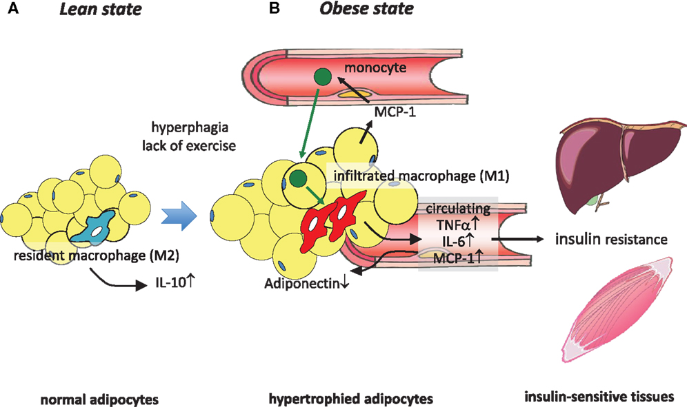
Let's look at this inflammatory cascade in more detail.
Adipocyte dysfunctions linking obesity to insulin resistance and type 2 diabetes
This scientific paper provides a fantastic step-by-step model of how this dysfunction progresses. We will focus on the figure and its description.
Please review Figure 1 and its detailed caption (section 3). Then, read the first two paragraphs of the text that follows (section 4). This will walk you through the progression from caloric excess to macrophage recruitment and the inflammatory state.
As described in the paper, this inflammatory environment, driven by cytokines like TNF-α, has a disastrous effect on adipocyte function. It impairs the fat cell's ability to store triglycerides and actually increases lipolysis (the breakdown of fat), further contributing to the "lipid spillover" of FFAs into the blood. It's a perfect storm of dysfunction.
Test your understanding!
A patient has obesity-related insulin resistance. Their blood tests show low levels of adiponectin and high levels of TNF-α. Based on what you've just learned, what does this hormonal profile tell you about the state of their adipose tissue?
Show answer
This profile is the classic signature of dysfunctional, inflamed adipose tissue. The low adiponectin indicates a loss of the healthy, insulin-sensitizing function of fat cells. The high TNF-α points to significant macrophage infiltration and chronic inflammation. This combination strongly suggests that the patient's adipose tissue is not just storing excess fat, but is actively promoting systemic insulin resistance through inflammatory pathways.
4. Synthesis: The Complete Picture
Let's now tie everything together. The relationship between obesity and insulin resistance is a chain of events rooted in adipose tissue dysfunction.
Obesity (Chronic Energy Surplus) ➡️ Adipose Tissue Expansion Limit Reached ➡️ Adipocyte Hypertrophy ➡️ Adipose Tissue Dysfunction, which includes:
- Impaired Lipid Buffering: Leads to lipid spillover (high FFAs) and ectopic fat deposition in muscle and liver.
- Chronic Inflammation: Leads to altered adipokine secretion (low adiponectin, high TNF-α, IL-6).
Both of these consequences directly cause insulin resistance in peripheral tissues. The free fatty acids and inflammatory cytokines interfere with insulin signaling pathways at the post-receptor level—exactly the mechanism we discussed in a previous lesson.

5. Metabolically Healthy Obesity (MHO): The Exception that Proves the Rule
To confirm that it's the dysfunction of fat, not just its quantity, that matters, we can look at a fascinating phenomenon known as Metabolically Healthy Obesity (MHO). These are individuals who are obese according to their BMI but do not display the expected signs of metabolic disease like insulin resistance, hypertension, or dyslipidemia.
What makes them different? Studies show that compared to their "metabolically unhealthy" counterparts with the same BMI, individuals with MHO tend to have:
- More effective fat storage in subcutaneous depots.
- Less visceral fat (fat around the internal organs).
- Smaller, more numerous adipocytes (hyperplasia over hypertrophy).
- Less ectopic fat in their liver and muscle.
- Lower levels of inflammation in their adipose tissue.
In short, their adipose tissue, while expanded, has remained largely functional.
Adipose tissue dysfunction in obesity
Let's return to the 'Adipose tissue dysfunction in obesity' video, where the speaker discusses the concept of MHO and presents data comparing healthy and unhealthy obese individuals.
Please watch from 08:05 to 12:05 and then from 15:13 to 17:19. The first part defines MHO and questions how 'healthy' it really is long-term. The second part presents key data showing that metabolically healthy obese individuals have smaller adipocytes and less macrophage infiltration in their fat tissue.
The existence of MHO powerfully reinforces our main thesis: it is the functional capacity and health of adipose tissue that dictates the metabolic consequences of obesity.
Conclusion
Today we have forged the crucial link between obesity and insulin resistance, completing our foundational understanding of the metabolic disturbances relevant to PCOS.
Key Takeaways:
- Adipose tissue is a vital endocrine organ, not just a passive storage site.
- The link between obesity and insulin resistance is adipose tissue dysfunction.
- This dysfunction is characterized by adipocyte hypertrophy (enlarged fat cells) when the capacity for healthy expansion (hyperplasia) is exceeded.
- Dysfunctional adipose tissue leads to two main problems:
- Lipid Spillover: Ectopic fat deposition in muscle and liver, causing local insulin resistance.
- Chronic Inflammation: Altered secretion of adipokines (low adiponectin, high TNF-α) that drives systemic insulin resistance.
- The concept of Metabolically Healthy Obesity (MHO) demonstrates that it is the function of fat tissue, not just its mass, that determines metabolic health.
Preview of the Next Module:
With this lesson, we conclude our deep dive into the fundamental physiology of hormones and metabolism. You now have a solid grounding in the HPO axis, the menstrual cycle, androgen physiology, and the intricate relationship between insulin resistance and obesity.
We are now perfectly positioned to apply this knowledge to our primary topic. Our next module, "PCOS: Definition, Diagnosis, and Epidemiology," will begin our formal study of the syndrome itself. We will start by defining PCOS, understanding its diagnostic criteria, and exploring its prevalence worldwide.