Hello! Welcome to the first lesson of our second module, "Female Reproductive Anatomy and Physiology."
In our previous module, we built a strong foundation in endocrinology, culminating in an understanding of how dynamic tests are used to probe the body's hormonal feedback loops. We saw that these tests are essential for diagnosing conditions like Cushing's syndrome or adrenal insufficiency, and even for distinguishing between PCOS and its mimics.
Now, we will pivot from the general principles of endocrinology to the specific organ at the heart of PCOS: the ovary. Today's lesson directly addresses the learning outcome: Describe the anatomical structure and cellular organization of the ovary, including follicles and stroma. We will dissect the ovary from its macroscopic structure down to its microscopic, cellular components. Understanding this anatomy is the critical first step before we can explore how its function is orchestrated during the menstrual cycle and how it becomes dysregulated in PCOS.
1. The Ovary: Macroscopic Anatomy
The ovaries are the female gonads, analogous to the testes in males. They are responsible for two primary functions: producing eggs (oocytes) and secreting hormones, mainly estrogen and progesterone.
To begin, let's get a clear picture of the ovary's overall structure and its main components.
The Female Reproductive System
This first reading from Lumen Learning's 'Anatomy and Physiology' provides a concise and clear overview of the ovary's location, its supporting ligaments, and its basic structural layers.
Please read the section titled 'Ovaries'. As you read, focus on identifying the three main regions of the ovary: The outer coverings: the ovarian surface epithelium and the tunica albuginea. The cortex: the outer portion containing the ovarian follicles. The medulla: the inner portion containing blood vessels, lymph vessels, and nerves.
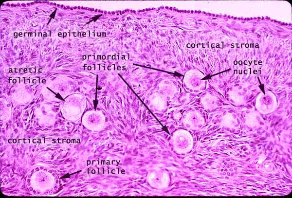
As you've just read, the ovary is not a uniform organ. It is organized into distinct functional zones. The cortex is the busy outer region where all the action of follicular development happens. The medulla serves as the vital support core, providing the blood supply and nerve connections necessary for the cortex to function. The connective tissue that makes up the bulk of the cortex is called the ovarian stroma. This stroma is not just inert packing material; it is a dynamic tissue that provides structural support and contains cells that will play a crucial role in hormone production.
2. Microscopic Anatomy: Ovarian Follicles
The fundamental functional units of the ovary are the follicles. Each follicle consists of an immature egg, called an oocyte, surrounded by one or more layers of supporting cells. The process of follicle growth and development is called folliculogenesis.
We will now take a detailed tour through the life stages of an ovarian follicle, from its most primitive state to a fully mature follicle ready for ovulation. Understanding these structures is essential, as the defining features of PCOS—such as "polycystic" ovaries and excess androgen production—originate from dysfunction at this microscopic level.
The following video provides an excellent and detailed histological walkthrough of the ovary, introducing each follicular stage.
Histology of the Ovary and Ovarian Follicles [Female Reproductive Histology Part 1 of 2]
The video 'Histology of the Ovary and Ovarian Follicles' from DaVinci Academy will guide us through the microscopic landscape of the ovarian cortex. It visually breaks down the complex process of folliculogenesis into distinct, identifiable stages.
Please watch the video from 00:21 to 15:52. As you watch, focus on identifying the key structural changes that define each stage of follicular development: (Starts at 00:21): The overall layers of the ovary (cortex, medulla, etc.). Primordial Follicle (starts at 06:12): Note the oocyte and the single layer of flattened follicular cells. Primary Follicle (starts at 07:19): Observe how the follicular cells become cuboidal (now called granulosa cells) and begin to multiply into layers (unilaminar vs. multilaminar). Note the appearance of the zona pellucida. Secondary (Antral) Follicle (starts at 10:59): See how the surrounding stroma organizes into the theca interna and theca externa. Notice the appearance of small, fluid-filled spaces. Mature (Graafian) Follicle (starts at 12:52): Watch as the fluid-filled spaces merge into a single large antrum. Identify the cumulus oophorus and corona radiata surrounding the oocyte.
3. A Summary of Follicular Stages
Let's consolidate what you've just seen. The development of a follicle is a continuum, but it's classified into stages based on clear structural milestones.

Here is a summary of the key features of each stage:
| Follicle Stage | Oocyte | Surrounding Cells | Key Features |
|---|---|---|---|
| Primordial | Primary Oocyte | Single layer of flat follicular cells | The most immature, resting stage. Forms the ovarian reserve. |
| Primary | Primary Oocyte | Single or multiple layers of cuboidal granulosa cells | Follicle "activates". Oocyte grows. Zona pellucida (a protective glycoprotein layer) forms around the oocyte. |
| Secondary | Primary Oocyte | Multiple layers of granulosa cells + Theca cells (interna & externa) | Stromal cells differentiate into theca layers. Small fluid pockets appear among granulosa cells. |
| Tertiary (Antral/Graafian) | Primary Oocyte (becomes Secondary Oocyte just before ovulation) | Granulosa cells + Theca cells | Fluid pockets merge to form a large, single cavity called the antrum. Oocyte is pushed to one side, surrounded by the cumulus oophorus and corona radiata. |
A crucial concept also mentioned in the video is atresia, the process by which most follicles degenerate and die. At any stage, a follicle can stop developing and undergo this programmed cell death. In a typical cycle, many follicles begin to grow, but only one becomes dominant and ovulates; the rest undergo atresia. In PCOS, this process is disrupted, leading to an accumulation of small follicles that have failed to either fully mature or undergo atresia—a state known as follicular arrest.
Test your understanding!
You are examining a histological slide of an ovary and see a follicle. It has multiple layers of cuboidal granulosa cells and a clearly defined layer of theca cells, but you do not see a single, large, fluid-filled antrum (though there might be tiny pockets of fluid).
How would you classify this follicle, and what is the key feature that prevents it from being classified as a mature Graafian follicle?
Show answer
This would be classified as a secondary follicle.
It is distinguished from a primary follicle because it has multiple layers of granulosa cells and has developed theca layers. However, the key feature it lacks to be a mature Graafian (tertiary) follicle is the presence of a single, large, confluent antrum. The development of this large fluid-filled cavity is the defining characteristic of the tertiary stage.
Conclusion
In this lesson, we have laid the anatomical groundwork for understanding ovarian function. We've moved from the macroscopic view of the ovary down to the intricate cellular architecture of its fundamental unit, the follicle.
Key Takeaways:
- The ovary is divided into an outer cortex, which contains the follicles and supportive stroma, and a central medulla with blood vessels and nerves.
- A follicle consists of an oocyte surrounded by supporting cells.
- Folliculogenesis is the process of development through distinct stages: primordial, primary, secondary, and tertiary (Graafian) follicles.
- Each stage is defined by specific structural changes, such as the shape of follicular cells, the formation of the zona pellucida, the development of the theca interna and externa, and the appearance of the antrum.
Preview of the Next Lesson:
Today, we focused on identifying the structures within the ovary. In our next lesson, we will build upon this by examining the process in more detail. We will address the learning outcome: "Explain the process of folliculogenesis from primordial follicle to mature Graafian follicle." This will involve looking at the timeline of this long journey, the factors that trigger a follicle to grow, and how a single follicle is selected to ovulate each cycle.