Hello! Welcome to the final lesson in our first module, "Foundations of Human Biology."
Introduction
In our last lesson, we took a deep dive into the cell's "powerhouse," the mitochondrion, and uncovered how it generates ATP, the energy currency that fuels all cellular activity. We saw that cells with high energy demands, like muscle and nerve cells, are packed with mitochondria.
This observation leads us to a broader concept: cells in the body are not all the same. They are specialized for different jobs. Today, we will move one level up in the body's hierarchy of organization, from individual cells to the structures they form when they work together. This lesson is all about tissues.
Your body is an incredibly complex organization, and much like in the business world, it relies on teamwork. If a cell is an individual worker, a tissue is a specialized team—like the finance department, the operations team, or the communications department—where groups of similar cells collaborate to perform a specific function. The study of these tissues is called histology.
By the end of this lesson, you will be able to identify the four main types of tissues in the human body and their functions. This knowledge will provide the framework for understanding how organs and entire organ systems are built and how they operate.
The Four Main Tissue Types
All the complex structures in your body are constructed from just four primary types of tissue:
- Epithelial Tissue (Covering)
- Connective Tissue (Support)
- Muscle Tissue (Movement)
- Nervous Tissue (Control & Communication)
Let's explore each of these "teams" to understand their unique roles.
1. Epithelial Tissue: The Protective Covering
Epithelial tissue, or epithelium, consists of tightly packed sheets of cells that cover surfaces and line body cavities. Think of it as the body's interface with its environment, both internal and external.
Key Functions:
- Protection: The epithelium of your skin protects your body from microbes, injury, and dehydration.
- Secretion: Glands, which are made of epithelial tissue, secrete substances like hormones, sweat, and digestive enzymes.
- Absorption: The lining of your small intestine is made of epithelial cells specialized for absorbing nutrients from food.
- Filtration: The epithelium in your kidneys helps filter your blood.
The video below gives a great overview of epithelial tissue, explaining how its structure (cell shape and layering) is perfectly suited to its function.
Tissues | Anatomy and Physiology | Comprehensive Overview!
This segment from the video 'Tissues | Anatomy and Physiology | Comprehensive Overview!' by Organized Biology explains what epithelial tissue is, how it's classified, and its main functions.
Please watch from timestamp 01:38 to 09:56. Pay attention to the link between the shape of the cells (flat, cube-like, column-like) and their primary jobs, like protection or secretion.
As the video explains, epithelial cells are packed together like bricks in a wall, creating a strong barrier. They have a distinct top (apical) side facing an open space and a bottom (basal) side anchored to the tissue underneath. A key point for our later discussions on health is that these cells divide rapidly to replace old or damaged ones, which is why a cut on your skin heals, but also why many cancers arise from this tissue type.
2. Connective Tissue: The Supporter and Connector
Connective tissue is the most abundant and diverse tissue type. As its name suggests, its primary role is to connect, support, bind, or separate other tissues and organs. Unlike the tightly packed epithelium, connective tissue cells are typically scattered within an extracellular matrix.
This matrix is the defining feature of connective tissue. It's composed of protein fibers (like strong collagen and flexible elastin) embedded in a "ground substance" that can be liquid (like in blood), gel-like (like in cartilage), or solid (like in bone).
You can think of the matrix using a Jello salad analogy: the cells are like pieces of fruit, the protein fibers are like shredded carrots, and the ground substance is the gelatin holding it all together.
The different types and arrangements of cells, fibers, and ground substance give rise to an amazing variety of tissues, including:
- Loose and Dense Connective Tissue: Found in ligaments, tendons, and the deep layers of your skin.
- Cartilage: Provides firm, flexible support in places like your joints, ears, and nose.
- Bone: Provides the rigid framework for your body.
- Adipose (Fat) Tissue: Stores energy, provides insulation, and cushions organs.
- Blood: A fluid connective tissue that transports gases, nutrients, and wastes throughout the body.
The following segments from the video "Introduction to Histology" walk through these different forms of connective tissue.
These clips from Dr. Morton's 'Introduction to Histology' provide a systematic look at the various types of connective tissue, from the 'proper' tissues to specialized forms like cartilage, bone, and blood.
Please watch from timestamp 18:56 to 28:34. Focus on understanding the general function of each type (e.g., bone for support, blood for transport) rather than memorizing all the cell names.
3. Muscle Tissue: The Mover
Muscle tissue is all about movement. It's composed of specialized cells that can contract, or shorten, to produce force. This ability allows you to walk, your heart to beat, and your stomach to churn food. There are three types of muscle tissue.
1. Skeletal Muscle:
- Function: Moves the skeleton.
- Control: Voluntary (you consciously control it).
- Appearance: Striated (has a striped or banded look under a microscope).
2. Cardiac Muscle:
- Function: Pumps blood.
- Control: Involuntary (contracts automatically).
- Appearance: Striated, and found only in the wall of the heart.
3. Smooth Muscle:
- Function: Controls movement in the walls of internal organs (e.g., stomach, intestines, blood vessels).
- Control: Involuntary.
- Appearance: Non-striated (smooth).
This next short clip clearly explains these three types.
This segment from 'Introduction to Histology' concisely describes the three types of muscle tissue, highlighting their differences in function, control, and appearance.
Please watch from 28:34 to 32:43.
4. Nervous Tissue: The Communicator
Nervous tissue is the body's command and control center. It is responsible for communication and coordination throughout the body. It forms the brain, spinal cord, and the vast network of nerves.
It consists of two main cell types:
- Neurons: These are the primary functional cells. They are specialized to generate and transmit electrical signals (nerve impulses), allowing for rapid communication between different parts of the body.
- Glial Cells (or Neuroglia): These are the "support staff" of the nervous system. They protect, insulate, and nourish the neurons.
The following clip provides a brief introduction to nervous tissue.
The final clip from 'Introduction to Histology' introduces the components of nervous tissue: neurons and their supporting glial cells.
Please watch from 32:43 to 37:11. This will set the stage for our next module, where we will explore the nervous system in detail.
Summary of Tissue Types
The image below provides a fantastic visual summary, pulling together the four tissue types with microscopic views and examples of where they are found in the body.
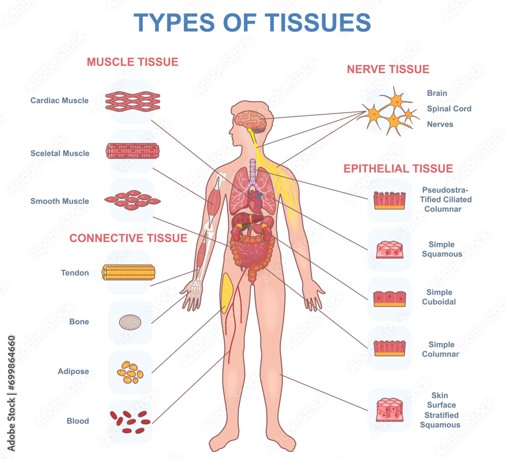
Here is a simple table to consolidate what we've learned:
| Tissue Type | Primary Function(s) | Key Characteristics | Examples |
|---|---|---|---|
| Epithelial | Covering, Protection, Secretion | Tightly packed cells in sheets | Skin, lining of digestive tract, glands |
| Connective | Support, Connection, Binding | Cells scattered in an extracellular matrix | Bone, cartilage, blood, fat, tendons |
| Muscle | Movement, Contraction | Elongated, contractile cells (fibers) | Skeletal muscles, heart, walls of organs |
| Nervous | Communication, Control | Cells generate and transmit electrical signals | Brain, spinal cord, nerves |
Test your understanding!
The wall of the stomach is an organ that has to withstand strong acid (protection), churn food (movement), and receive signals from the brain (control). Which of the four main tissue types would you expect to find performing each of these three functions?
Show answer
- Epithelial tissue would line the stomach to protect the underlying layers from acid and to secrete mucus and digestive juices.
- Muscle tissue (specifically, smooth muscle) would be in the walls to contract and churn the food.
- Nervous tissue would be present to control the muscle contractions and secretions, and to send signals (like fullness or pain) back to the brain.
- (Bonus: Connective tissue would also be there, binding all these layers together and supplying them with blood!)
Conclusion
Congratulations on completing the first module! We've built a solid foundation, starting from the body's overall organization and moving down to the cellular level to see how energy is made, and now back up to see how cells team up to form tissues.
Here are the key takeaways from this lesson:
- A tissue is a group of similar cells that work together to perform a specific function.
- There are four main types: epithelial (covers and protects), connective (supports and binds), muscle (moves), and nervous (communicates).
- The structure of each tissue type is directly related to its function. For example, the tightly packed nature of epithelial tissue makes it a great barrier, while the contractile fibers of muscle tissue make it ideal for generating force.
In our next module, we will begin our journey through the major organ systems. We'll start with the body's master control system by taking a closer look at the nervous tissue we just introduced. Our first lesson will identify the major divisions of the nervous system—the Central and Peripheral—and their primary roles in running your body.