Hello. Last lesson, you traced the handoff at a chemical synapse: an arriving action potential triggers neurotransmitter release, and receptors on another cell produce a response. This final lesson in the foundations module asks you to use that mechanism as a diagram-checking tool. You’ll distinguish a chemical synapse from an electrical synapse, then see how to correct annotations that confuse the two. Allow about 35–40 minutes, including time to inspect the figure closely.
What crosses between the cells?
The word electrical can cause a misleading first impression. Electrical activity occurs in both kinds of synapse: an action potential can arrive at a chemical synapse, and the receiving cell can undergo a voltage change. The distinction is how the signal passes between cells.
At a chemical synapse, the cells are separated by a synaptic cleft. An action potential at the sending terminal opens voltage-gated calcium channels; calcium enters that terminal and helps trigger vesicle fusion. Released neurotransmitter molecules diffuse across the cleft and bind receptors on the receiving cell. A postsynaptic voltage change is one possible result.
At an electrical synapse, protein channels called gap junctions connect the interiors of adjacent cells. Ions can pass directly between them, carrying current without the vesicle-release and receptor-binding steps required for chemical transmission. That current can change the neighbouring cell’s voltage; it does not guarantee that the neighbouring cell fires an action potential.
Watch the short comparison below for the gap-junction structure. Concentrate on the physical route taken by the ions, rather than treating “electrical” as a synonym for “faster action potential.”
10-Minute Neuroscience: Synapses
Watch “10-Minute Neuroscience: Synapses” from Neuroscientifically Challenged to see how a gap junction connects two cells.
In the “electrical synapses” portion, watch gap junctions. Follow the channels bridging the two membranes and the ions moving between cells. Notice that the discussion describes rapid transmission and often two-way current flow; neither means that every electrical synapse works identically.
For a written comparison, read the “Electrical synapses” subsection of Khan Academy’s The synapse. Its note that some synapses have both electrical and chemical components is a useful guard against forcing every real connection into an exclusive category.
The synapse (article) | Human biology
Read Khan Academy’s comparison to check what gap junctions permit and why chemical synapses offer different signalling possibilities.
Under “Electrical synapses,” begin at the paragraph starting “At electrical synapses, unlike chemical synapses.” Read the comparison through the final paragraph before “References.” Inspect the electrical-synapse figure, and note the qualifications “in many cases” for two-way current flow and “some synapses” for connections with both forms of transmission.
Read the anatomy before trusting the labels
The following figure places the two mechanisms side by side. In panel a, vesicles contain green neurotransmitter molecules; the molecules are released between the cells and encounter receptors on the lower cell. In panel b, blue gap-junction channels bridge the two cells. The arrows inside those channels depict possible directions of current flow.
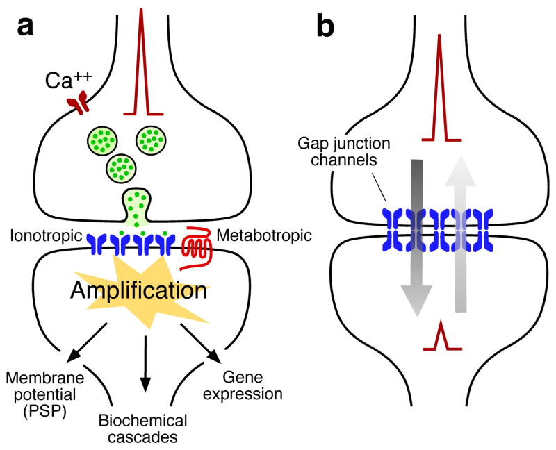
When inspecting an annotated diagram, locate the two membranes first. Then trace each labelled substance through a specific route. In panel a, calcium enters the upper terminal from outside it. Neurotransmitter leaves that terminal and crosses the space between the cells. If a receptor subsequently changes an ion channel’s state, ions cross the receiving cell’s own membrane. Those are three distinct movements. Labelling all of them “ions crossing the synapse” would erase the mechanism you need to explain.
Panel a also shows two kinds of postsynaptic receptor. An ionotropic receptor is itself an ion channel whose state changes when a messenger binds. A metabotropic receptor works indirectly through signalling inside the cell. You do not need its intracellular details yet. For diagram checking, the point is that a postsynaptic response need not be direct channel opening, and neither receptor type guarantees a new spike.
Panel b has no vesicle-release route drawn. The blue structures join the cells through gap-junction channels, allowing ions to move from one cell’s interior to the other’s. The two red spikes are an illustration of activity in both cells, not a rule that one arriving spike must produce an identical spike in its neighbour. Depending on the junction and the cells’ electrical states, current can often flow in either direction. In that situation, “sender” and “receiver” describe the direction at a particular moment rather than permanent roles.
A worked diagram correction
Suppose someone has annotated a copy of panel a with this sentence: “The action potential crosses the synaptic cleft through the calcium channel and opens an identical channel on the next neuron.”
The first repair is location. The calcium channel belongs in the presynaptic terminal membrane, not across the cleft. The second is identity: calcium enters the terminal, while neurotransmitter molecules cross the cleft. The third is consequence: receptors on the receiving cell may change its activity, but they do not automatically make it fire an identical action potential. A corrected annotation would read:
“An action potential reaches the upper terminal. Calcium enters through channels in that terminal’s membrane and helps trigger neurotransmitter release. Neurotransmitter crosses the cleft and binds receptors on the lower cell, producing a postsynaptic response.”
Now suppose an annotation on panel b reads: “Vesicles release neurotransmitter into the gap junction; receptors then pass ions into the second neuron.” Here the objects have been taken from the other panel. A gap junction is a channel connecting the two cell interiors, not a container for neurotransmitter or a receptor that waits for its release. The corrected annotation is:
“Gap-junction channels directly connect the cells, allowing ions to carry current between them and alter the neighbouring cell’s voltage.”
These examples reveal a reliable way to diagnose an unfamiliar annotation. Check what moves, where it moves, and what the movement can establish. A label may name a real biological object yet still be wrong because it puts that object on the wrong membrane or assigns it the wrong job. A final check is the strength of the claim: replacing “may change the receiving cell’s voltage” with “always produces a spike” introduces an error even when the structures are labelled correctly.
The contrast is useful, but it is not an absolute division between “chemical neurons” and “electrical neurons.” A neuron can participate in different kinds of connections, and some junctions have both electrical and chemical components. Likewise, chemical synapses commonly transmit from a releasing terminal to a receiving cell, while electrical junctions often permit current in either direction; these are descriptions of typical mechanisms, not permission to ignore the structures shown.
The distinction to carry forward
To correct a synapse diagram, do more than swap the headings chemical and electrical. At a chemical synapse, look for presynaptic calcium entry, vesicle release, neurotransmitter crossing a cleft, and postsynaptic receptor action. At an electrical synapse, look for gap-junction channels linking the cells and direct ion-mediated current flow between them. In either case, distinguish an effect on the receiving cell’s voltage from a guaranteed new action potential.
You now have the cellular framework needed for the next module. Its first lesson follows a particular neurotransmitter—dopamine—beginning with how a neuron synthesizes it before packaging and releasing it.