Recommended resources
The freely available resources this lesson will direct you to.
- KS3 Biology - Animal & Plant Cellswww.youtube.com
- MICROGRAPHS | The secret to identifying organelleswww.youtube.com
- Plant cells - The key features of cells and their functions - OCR 21st Century - GCSE Biology (Single Science) Revision - Cambridge OCR 21st Century - BBC Bitesizewww.bbc.co.uk
- Learn and try: Cell parts and functions (article) | Khan Academywww.khanacademy.org
Hello! Last time, you connected cell structures to their functions. You also saw the features commonly associated with plant cells: a cell wall, chloroplasts, and a large central vacuole. This lesson changes the task from explaining those structures to using visible evidence to identify a cell.
That matters in an MYP eAssessment because an image may be an unfamiliar drawing or a photograph taken through a microscope. The strongest answer does not rely on a cell looking “plant-shaped”; it identifies features you can actually see and recognises what the image may be too unclear to show. Allow about 35–40 minutes, including time to inspect the images.
Start with a diagram—but do not mistake it for a photograph
Look at the two cells below. In the plant cell, find the thick cell wall outside the thinner cell membrane, the green chloroplasts, and the large, pale central vacuole. Then find the nucleus and mitochondria in both cells.
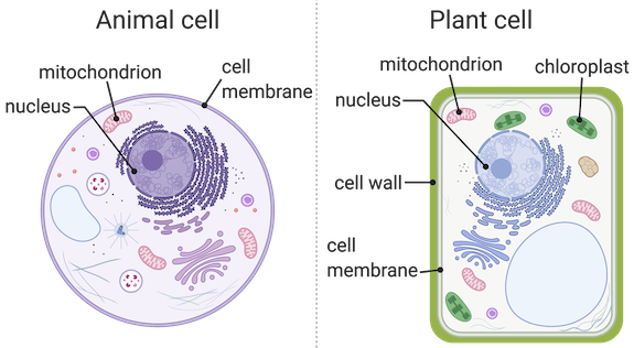
The most useful clue in this drawing is the cell wall. It is an additional boundary outside the plant cell’s membrane; the animal cell has a membrane but no wall. Chloroplasts are also decisive evidence for a plant cell when they are clearly identified. A large central vacuole adds support to the identification, but the small pale space in the animal-cell drawing shows why “has a vacuole” is not enough on its own.
The nucleus and mitochondria are different kinds of clues: they tell you something about what is inside the cells, but they do not distinguish plant from animal here. Nor does colour alone. Diagram-makers choose colours to make parts easy to recognise; real cells will not necessarily have those colours in a microscope image.
Before moving on, trace each cell’s outer edge with your eye. In the plant cell, there are two boundaries to distinguish: wall outside, membrane just inside. In the animal cell, the membrane forms the outer boundary. That distinction will become harder to see in a micrograph.
What changes when you look at a micrograph?
A diagram is a model whose details have been selected and drawn clearly. A micrograph is an image of real cells captured using a microscope. Its features depend on the specimen, preparation, and what the microscope can resolve. Study the four-image comparison in the following reading with that difference in mind.
Learn and try: Cell parts and functions (article) | Khan Academy
Read Khan Academy’s comparison of cell diagrams and micrographs to see why a labelled model is easier to interpret than an image of real cells.
In “Cell diagrams represent real cells,” start with the sentence “Cells are too small to be seen with the naked eye.” Read the comparison through the caption beneath the four images labelled A–D; stop before “Now, let's take a closer look.” Compare animal diagram A with blood-cell micrograph B, then plant diagram C with plant-cell micrograph D. Notice which features remain recognisable and which are no longer obvious.
The comparison reveals a trap: an organelle missing from your view is not necessarily missing from the cell. Ribosomes are far too small to pick out in an ordinary light micrograph, and the membrane may be difficult to distinguish where it lies close against a plant cell wall. A nucleus may also be difficult to see, depending on the specimen. Mature mammalian red blood cells, such as those shown in the reading, are an additional exception: they lack a nucleus.
It helps to separate absence of evidence from evidence of absence. “I cannot see a chloroplast” is only a statement about the image. It does not prove that the cell is animal: some plant cells, including many cells in roots, do not have chloroplasts.
Read the boundaries first in an unfamiliar micrograph
When a micrograph has no helpful labels, begin at the cell’s edge rather than guessing from its overall shape. Plant cells often show a relatively firm, conspicuous boundary. In a group of plant cells, walls may appear as lines separating neighbouring cells. An animal cell has a membrane but no rigid cell wall. Its boundary may appear thinner or less regular, although tightly packed animal cells can also form an orderly pattern.
Next, look within the boundary. Depending on the specimen, chloroplasts may appear as several small, darker bodies in the cytoplasm. A large central vacuole may appear as a broad, paler region that leaves the cytoplasm nearer the edge. Neither feature is guaranteed to be clear in every image. In particular, do not label every dark oval a chloroplast: a nucleus may also appear as a darker body, and its position and appearance will vary.
This short micrograph demonstration lets you see the boundary and internal features being identified in photographs. Its images are more detailed than many school light-microscope images, so concentrate on the method of looking, not on memorising every visible organelle.
MICROGRAPHS | The secret to identifying organelles
Watch selected parts of Miss Angler’s “MICROGRAPHS | The secret to identifying organelles” to see how the appearance of a cell differs from a tidy diagram.
Watch plant boundaries for the outer wall and the much harder-to-see membrane. Then watch internal clues for examples of chloroplasts and a vacuole; pause where useful to locate them yourself. Finish with animal-cell features, focusing on the outer boundary and nucleus. You do not need to identify the smaller organelles discussed elsewhere in the video to decide whether a cell is plant or animal.
Notice why shape is supporting evidence, not a rule. A regularly arranged group of cells with visible walls strongly suggests plant tissue. Yet an individual plant cell need not be a perfect rectangle, and an animal cell need not be round. Features at the boundary and inside the cell make a stronger case than its outline alone.
Build an evidence-based identification
Suppose a micrograph shows rows of cells divided by clear, firm-looking boundaries. Within each cell is a broad pale area, but no chloroplasts are visible. The best identification is plant cells: the boundaries provide evidence of cell walls, and the pale areas are consistent with large vacuoles. The lack of visible chloroplasts does not overturn that evidence.
Now suppose a different image shows cells with visible nuclei and thin outer boundaries, but no clear walls or chloroplasts. These features are consistent with animal cells. How confidently you can identify them depends on image quality: if the edges are blurred, you should not claim that you have proved a wall is absent. A nucleus alone cannot settle the question because plant cells can have nuclei too.
For an assessment response, match the strength of your wording to the strength of the image. If a wall or chloroplasts are clear, you can give a firm identification and name the visible evidence. If the image is ambiguous, say which type it most likely shows and what feature prevents certainty. This is more precise than listing everything you cannot see.
A useful way to organise your inspection is:
- Boundary: Is there evidence of a cell wall outside the membrane?
- Interior: Are chloroplasts or a large central vacuole visible?
- Limits: Could an absent-looking feature simply be hidden, unresolved, or not present in this particular plant cell?
Key takeaways
To distinguish plant from animal cells, give greatest weight to a visible cell wall or clearly identified chloroplasts. A large central vacuole can strengthen a plant-cell identification; a nucleus or mitochondrion cannot distinguish the two. In a micrograph, some structures may be unclear or invisible, so do not treat “I cannot see it” as proof that it is absent.
Next, you’ll use the same habit of examining structure as evidence to infer what a specialised cell does.