Recommended resources
The freely available resources this lesson will direct you to.
- Bone density : in implant | misch classificationwww.youtube.com
- Dental Implants 101: What You NEED to Know! Part 3 (Everything Bone Grafting)www.youtube.com
- Bone mineral density as a criterion for primary implant...journals.lww.com
- MINI-REVIEW REVISITING BONE DENSITY - MM Publisherswww.editorialmanager.in
Hello! Welcome to the final lesson of our second module.
In our previous lesson, we detailed the biological cascade of fracture healing, understanding how bone repairs itself through a sequence of inflammatory, reparative, and remodeling phases. We established that the mechanical environment—specifically the amount of strain at the injury site—dictates whether healing is direct or indirect.
Today, we build directly on that foundation. The success of the healing process, and by extension, the success of a dental implant, is not just a function of the biological pathway but is critically dependent on the quality of the material that is healing: the bone itself. This lesson addresses the learning outcome: Explain the clinical significance of bone quality and density (e.g., D1-D4 classification) for implant success.
We will explore how bone is classified based on its internal architecture, where these different types of bone are typically found in the jaws, and how clinicians assess this quality. Most importantly, we will connect bone density to the crucial concept of primary stability, which influences everything from surgical technique to the long-term prognosis of the implant.
1. Classifying Bone: The Misch D1-D4 System
Just as materials in engineering are characterized by their properties, bone can be characterized by its quality and density. This refers to its internal architecture—specifically, the ratio and arrangement of dense cortical bone and porous trabecular (or cancellous) bone.
The most widely used system for this was developed by Carl Misch, who categorized bone into four types, from most dense (D1) to least dense (D4).
To start, let's get a clear overview of this classification.
Bone density : in implant | misch classification
The video 'Bone density : in implant | misch classification' provides a concise introduction to the four bone density types and their typical locations in the jaw.
Please watch from the beginning until 07:05. The video will cover: The importance of bone density for biomechanical properties (0:00 - 1:34). How density varies by location (mandible vs. maxilla) (1:34 - 4:34). A description of each of the four Misch classifications (D1-D4) (4:34 - 7:05).
The video provides a great summary. Here is a visual representation of the four bone types described.

To solidify this, let's turn to a review paper that details these categories.
REVISITING BONE DENSITY: A REVIEW ON MISCH CLASSIFICATION
The paper 'REVISITING BONE DENSITY: A REVIEW ON MISCH CLASSIFICATION' offers a more detailed textual description of the bone types and their prevalence in different parts of the jaws.
Read the 'Introduction' and the section on 'Location'. This will reinforce the macroscopic structure of each bone type and provide more specific data on where you are likely to find D1, D2, D3, or D4 bone.
As the resources explain, the general distribution is:
- Mandible (Lower Jaw): Generally denser, due to its function as a force-bearing, independent structure. The anterior mandible is often D2, while the posterior can be D2 or D3.
- Maxilla (Upper Jaw): Generally less dense and more porous. The anterior maxilla is often D3, while the posterior maxilla, particularly near the sinuses, is frequently D4.
This anatomical variation in bone density is a primary reason why implant success rates have historically been higher in the mandible than in the maxilla.
2. Assessing Bone Density
Clinicians have several ways to determine the bone density at a potential implant site.
Tactile Sensation
During the osteotomy (drilling) procedure, an experienced surgeon can get a "feel" for the bone's density. The resistance encountered by the drill provides a direct, albeit subjective, assessment.
The review paper you just read offers a useful analogy for this tactile feedback.
REVISITING BONE DENSITY: A REVIEW ON MISCH CLASSIFICATION
Please return to the 'REVISITING BONE DENSITY' paper and read the section on tactile sense.
Read the section titled 'Tactile Sense'. It compares the sensation of drilling into the four bone types to drilling into different densities of wood and other materials.
This analogy provides an intuitive physical understanding:
- D1: Like drilling into oak or maple (hardwood).
- D2: Like drilling into pine (softwood).
- D3: Like drilling into compressed balsa wood.
- D4: Like drilling into styrofoam.
Quantitative Assessment: CBCT and Hounsfield Units
While tactile sense is useful, a more objective, quantitative assessment is needed for pre-surgical planning. This is achieved using Cone Beam Computed Tomography (CBCT).
A CBCT scan produces a 3D image of the jaw. The radiodensity of the tissue in each voxel of the scan is quantified on a grayscale. This can be expressed in Hounsfield Units (HU), a scale originally developed for conventional medical CT. The HU scale is a linear transformation of the underlying X-ray attenuation coefficient, where air is defined as -1000 HU and water is 0 HU. Denser tissues have higher HU values.
The following research paper establishes the critical link between these quantitative HU values, the D1-D4 classification, and implant outcomes.
Bone mineral density as a criterion for primary implant...
The paper 'Bone mineral density as a criterion for primary implant...' is a clinical study that quantifies the relationship between bone density and implant stability. It provides the evidence base for the clinical significance we are discussing.
First, read the 'INTRODUCTION' section. It explicitly links the D1-D4 classification to ranges of Hounsfield Units. Then, read the subsection 'Cone beam computed tomography scan' to see how these measurements are practically made.
As the paper outlines, the Misch classification can be correlated with HU ranges:
- D1 bone: > 1250 HU
- D2 bone: 850–1250 HU
- D3 bone: 350–850 HU
- D4 bone: 150–350 HU
This ability to quantify bone density before surgery is a cornerstone of modern implant planning.
3. The Clinical Significance of Bone Density
Now we arrive at the central question: why does this classification matter so much? Bone density directly impacts implant success through its effect on primary stability.
Primary stability is the initial mechanical fixation of the implant in the bone immediately after placement. It is a measure of the absence of mobility. This initial stability is arguably the most critical factor for successful osseointegration.
Primary Stability and Healing
Recall from our last lesson that low strain (<2%) promotes direct bone healing (intramembranous ossification), while higher strain can lead to cartilage or fibrous tissue.
- High Primary Stability (Dense Bone, D1/D2): The implant is tightly gripped by the bone, resulting in very low micromotion under load. This creates the ideal low-strain environment for direct bone healing at the implant surface, leading to predictable and rapid osseointegration.
- Low Primary Stability (Soft Bone, D4): The implant has less initial grip. If micromotion exceeds a critical threshold (typically cited as 50-150 µm), the healing response shifts away from bone formation. Instead, a layer of fibrous connective tissue forms around the implant—a state known as fibrous encapsulation, which is a clinical failure.
The paper you've been reading provides direct, quantitative evidence for this link. It correlates the pre-operative bone density (in HU) with the insertion torque—a clinical measure of the rotational force required to seat the implant, which serves as a proxy for primary stability.
Bone mineral density as a criterion for primary implant...
Let's examine the results of the study. Please return to the paper 'Bone mineral density as a criterion for primary implant...'
Read the 'RESULTS' and 'DISCUSSION' sections. Focus on the correlation between bone density (HU), insertion torque (Ncm), and implant success/failure rates. Note the significant difference in mean HU and torque values between successful and failed implants.
The study's findings are clear: successful implants were placed in bone with significantly higher mean density and achieved higher mean insertion torque compared to failed implants. The majority of failures occurred in the posterior maxilla, the region most associated with D4 bone.
Adapting the Treatment Plan to Bone Density
Because bone density is so critical, it dictates nearly every aspect of the treatment plan:
- Surgical Technique: In soft D4 bone, the surgeon may use a narrower drill sequence to intentionally under-prepare the osteotomy. This causes the implant to compress the surrounding bone as it's inserted (osseodensification), increasing primary stability.
- Implant Design: Implants designed for softer bone often have more aggressive thread designs, a more pronounced taper, or a greater surface area to engage more of the available bone.
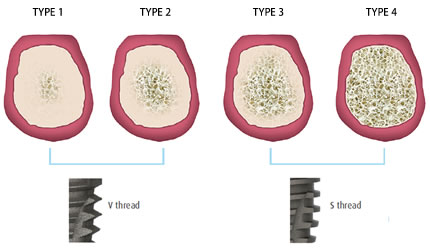
- Healing Time and Loading Protocol:
- In dense D1/D2 bone with high primary stability (>35 Ncm torque), a clinician might consider immediate loading, where a temporary crown is placed shortly after surgery.
- In soft D4 bone with low primary stability, a two-stage, submerged protocol is standard. The implant is covered by the gums and allowed to heal for an extended period (4-6 months or more) without any load before a crown is attached.
The concept of "biotype" (thin vs. thick bone and gums) is also related, as it affects the long-term stability and aesthetics around the implant.
Dental Implants 101: What You NEED to Know! Part 3 (Everything Bone Grafting)
This video discusses the related concept of bone 'biotype', which reinforces the idea that the inherent quality and quantity of a patient's tissue are fundamental to success.
Watch the segment from 13:51 to 16:24. It visually demonstrates the difference between a patient with a 'thick biotype' (robust bone) and a 'thin biotype' (delicate bone) using CBCT scans.
Conclusion
We have now completed our module on the biological foundations of bone. By understanding bone's structure, its dynamic remodeling, its healing process, and its varying quality, you have built a comprehensive picture of the biological environment into which a dental implant is placed.
Key Takeaways:
- Bone quality is categorized using the Misch D1-D4 classification, which describes the macroscopic ratio of cortical to trabecular bone.
- Bone density is not uniform; the mandible is typically denser (D2/D3) than the maxilla (D3/D4), with the posterior maxilla being the least dense region.
- Clinicians assess bone density through tactile sensation during surgery and, more importantly, through pre-operative CBCT scans, which provide quantitative Hounsfield Unit (HU) values.
- Bone density is clinically significant because it determines primary stability. Denser bone provides a better initial grip, promoting direct bone healing and successful osseointegration.
- Treatment planning, including surgical technique, implant selection, and loading protocols, must be adapted to the site-specific bone density to mitigate risks and ensure predictable outcomes.
Preview of the Next Lesson:
Having thoroughly examined the "host" (the bone), we will now turn our attention to the "guest" (the implant). In the first lesson of our next module, "Implant Materials and Biomechanical Design," we will address the learning outcome: Explain the concept of biocompatibility and the specific material properties of titanium and its alloys (e.g., elastic modulus, corrosion resistance, surface passivation) that enable osseointegration. We will investigate what makes titanium the material of choice for creating a direct, lasting bond with living bone.