Recommended resources
The freely available resources this lesson will direct you to.
Hello! Let's continue our exploration of decompression theory.
Introduction
In our last lesson, we established that supersaturation—the state where the inert gas pressure in your tissues exceeds the ambient pressure—is the fundamental driver of decompression stress. We concluded that the goal of any decompression strategy is to manage this supersaturation, keeping it below a "tolerable limit" to prevent symptomatic bubble formation.
Today, we will give that "tolerable limit" a name and a number. This lesson focuses on two of the most important concepts in modern decompression planning: M-values and gradient factors. You will learn:
- What M-values are and how they define the absolute limits of a dissolved-gas decompression model.
- What gradient factors are and how they provide a powerful tool for you, the diver, to control the conservatism of your decompression profile.
By the end of this lesson, you will understand the core mechanics of how your dive computer calculates a decompression schedule and how you can tailor it to your personal risk tolerance and the specific conditions of a dive.
Part 1: M-Values - Defining the Limit
The concept of a tolerable limit of supersaturation was first quantified by Robert Workman in the 1960s and later refined by Dr. Albert A. Bühlmann. They introduced the term M-value, where 'M' stands for "Maximum".
An M-value is the maximum inert gas pressure (often called tension) that a theoretical tissue compartment can withstand at a given depth (ambient pressure) without forming symptomatic bubbles.
Each tissue compartment in the model (e.g., the 16 compartments in Bühlmann's ZHL-16 model) has its own unique M-value.
Decompression Theory for the PADI RDP exam – M-values
To start, let's watch a video that provides a clear, conceptual overview of M-values and their role in no-stop diving.
Watch the section from 00:49 to 03:26. Focus on the definition of an M-value, its relationship to no-stop limits, the inverse relationship between half-times and M-values (faster tissues generally have higher M-values), and the concept of a 'controlling compartment'.
As the video explains, the M-value isn't a single number. It changes with depth. The greater the ambient pressure, the more supersaturation a tissue can tolerate. This relationship is linear and can be described by a simple equation for each compartment. This means we can plot the M-value for each compartment as a line on a graph of pressure vs. depth.
Diving Theory: What are M-values?
This article from Dive-Scuba.com provides a more detailed look at M-values, including how they are calculated and visualized.
Please read the sections titled 'What are M-values?' and 'How are M-values calculated?'. Pay close attention to the graphs that show the M-value as a line that changes with depth. The final graph showing all 16 M-value lines is particularly important.
From the reading, here are the key points to consolidate:
- M-values are depth-dependent: They increase linearly with ambient pressure.
- Each compartment has a unique M-value line: Slower tissues are less tolerant of supersaturation and thus have lower M-values than faster tissues at any given depth.
- The M-value line is the "hard deck": In a pure Bühlmann model, your decompression ceiling is the depth at which your leading compartment's inert gas pressure equals its M-value. Ascending shallower than this ceiling is predicted to cause an unacceptable risk of DCS.
Part 2: Gradient Factors - Controlling Your Conservatism
Diving at the M-value is like driving at the absolute edge of the road. It's the theoretical limit, but it leaves no room for error or for individual factors like cold, exertion, or predisposition to DCS. Technical divers needed a way to build a programmable safety margin into the model. This led to the development of gradient factors (GF) by Erik Baker.
Gradient Factors allow a diver to create a new, more conservative decompression ceiling, which is a set percentage of the original M-value. They are expressed as two numbers: GF Low and GF High.
SCUBA SCIENCE 10: Decompression Theory Explained Simply | Gradient Factors, Bühlmann, RGBM
This video from Dive SAGA gives a concise explanation of how GF Low and GF High work to shape your decompression profile.
Watch the segment from 10:01 to 11:36. The key is to understand what the 'low' number controls and what the 'high' number controls.
Let's break this down with a more detailed explanation.
- GF Low (GF Lo): This determines the conservatism of your first decompression stop. It's expressed as a percentage of the M-value at that depth. A lower GF Lo forces a deeper first stop, giving slow tissues more time to off-gas before you reach shallower depths.
- GF High (GF Hi): This determines the conservatism at the surface. It's a percentage of the surface M-value and dictates how much residual gas pressure you're willing to tolerate upon surfacing. A lower GF Hi means you will be "cleaner" (have less residual nitrogen) at the end of your dive.
The dive computer then creates a sloping ceiling that transitions from your GF Low percentage at the first stop to your GF High percentage at the surface.
The Dive Rite website has an excellent article that explains this concept in detail. It's one of the best resources for understanding GFs.
Please read the section titled 'GRADIENT FACTORS'. It clearly explains the concept of GF 0% and GF 100% and then introduces the GF Low/High method.
The following images are crucial for visualizing how this works.
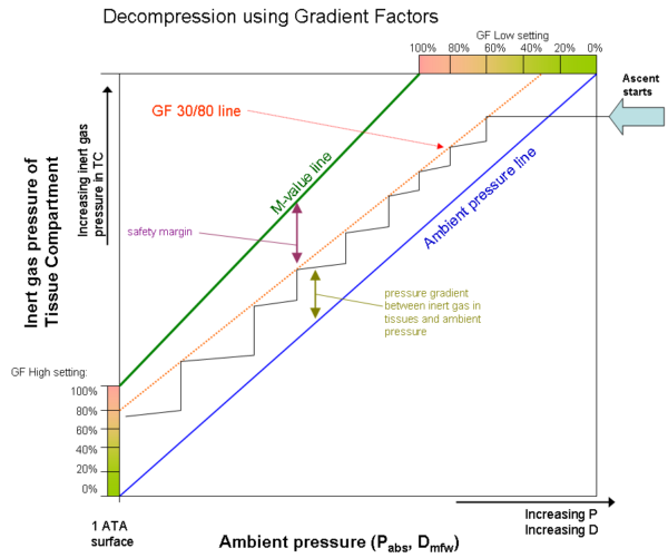
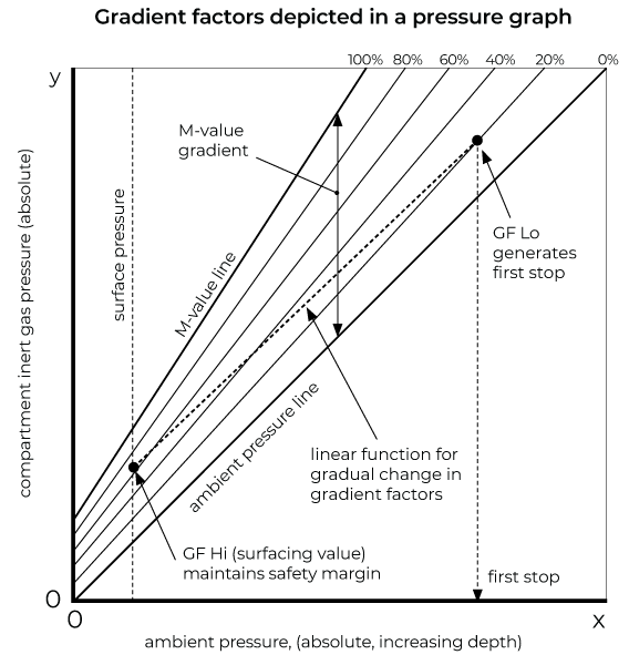
A common setting for technical diving might be 30/85. This means:
- The diver's first decompression stop will be at a depth where the leading tissue compartment's pressure reaches 30% of the M-value margin.
- The ascent will proceed along a sloping ceiling that gradually allows more supersaturation, up to 85% of the M-value margin.
- The diver will surface only when all compartments are below 85% of their respective surface M-values.
Part 3: Practical Implications
Choosing your gradient factors is one of the most important decisions in technical dive planning. Different settings can dramatically alter your decompression profile, total dive time, and gas requirements.
- Conservative settings (e.g., 30/70): Result in deeper first stops and longer overall decompression times. This is often used for deep, cold, or strenuous dives where you want to minimize decompression stress.
- Aggressive settings (e.g., 50/90): Result in shallower stops and shorter decompression. These might be used by a diver who is well-rested, warm, and doing a less demanding dive, but they carry a higher statistical risk of DCS.
- "Standard" settings (e.g., 40/85): A common starting point that balances safety with efficiency.
It's crucial to understand that there is no single "correct" GF setting. The choice is a form of personal risk management. It's your responsibility as a diver to understand what these numbers mean and to select settings that are appropriate for you and for the specific dive you are planning. Copying another diver's settings without understanding the rationale is a dangerous practice.
Conclusion
Today, we have demystified the core of modern decompression algorithms. You now understand the concepts that govern your dive computer's calculations.
Key Takeaways:
- M-values are the maximum tolerated inert gas pressures for each tissue compartment at a given depth. They represent the absolute limit of the Bühlmann model.
- Gradient Factors (GF) are a user-defined tool to apply a safety margin to the M-values, creating a more conservative decompression ceiling.
- GF Low controls the depth of the first stop, influencing the deep phase of decompression.
- GF High controls the allowable supersaturation upon surfacing, influencing the shallow phase and your post-dive condition.
- The choice of GF settings is a critical part of dive planning and risk management.
Preview of the Next Lesson:
The M-value and gradient factor system we've discussed is the heart of dissolved gas models (like Bühlmann's). However, this is not the only approach to managing decompression. In the next lesson, we will compare and contrast this approach with bubble models (such as VPM and RGBM), which are based on different fundamental assumptions about how bubbles form and grow in the body.