Welcome back. In our previous lesson, we examined the specialized cells that make up bone tissue. We learned that osteoblasts are the "builders," responsible for creating new bone matrix. Now, we'll see these cellular builders in action as we explore the two fascinating processes by which the human skeleton is constructed from scratch.
This lesson focuses on comparing intramembranous and endochondral ossification. These are the two pathways for bone formation, or osteogenesis, that occur during fetal development. Understanding these distinct mechanisms is crucial, as they explain why different bones in the body develop differently. For your nursing studies, this knowledge provides the foundation for understanding developmental skeletal abnormalities, how fractures heal (which often recapitulates these processes), and the significance of a baby's "soft spots."
An Overview of Bone Formation
Before we dive into the details, let's get a high-level overview. All bone begins with embryonic connective tissue called mesenchyme. From there, the paths diverge. The core difference lies in whether bone forms directly from this mesenchyme or if it first creates a cartilage "model" that is later replaced by bone.
To get a quick and clear sense of this fundamental distinction, let's watch a brief video.
Intramembranous vs. Intracartilagenous Ossification (Endochondral) 🦴 - Anatomy Series
This short video from Medicosis Perfectionalis provides a very concise and energetic comparison of the two ossification types.
Watch the video from the beginning to the end. Focus on the key concepts of a direct versus an indirect process and which one is faster.
As the video highlights, the two processes are:
- Intramembranous Ossification: A direct process where bone develops from a fibrous membrane of mesenchymal cells. It's faster and forms the flat bones of the skull, parts of the clavicles, and the mandible.
- Endochondral Ossification: An indirect process where a hyaline cartilage model is created first and then gradually replaced by bone. This is a slower, more complex process responsible for forming most bones in the body, especially long bones.
This diagram gives you a clear visual map of which bones in the skeleton are formed by each method.

Now, let's explore each of these pathways step-by-step.
Intramembranous Ossification: Direct Bone Formation
The term "intramembranous" means "within a membrane." This process is how bone develops directly from sheets of mesenchymal connective tissue. It's the simpler of the two pathways.
The following reading from OpenStax "Anatomy & Physiology 2e" provides an excellent, illustrated guide to the four main steps of this process.
6.4 Bone Formation and Development - Oregon State University
This text describes the sequence of events in intramembranous ossification, accompanied by a very helpful diagram.
Please read the section describing the process. As you read, follow along with the four steps shown in Figure 6.4.1 (also shown below) to visualize how the bone tissue develops.
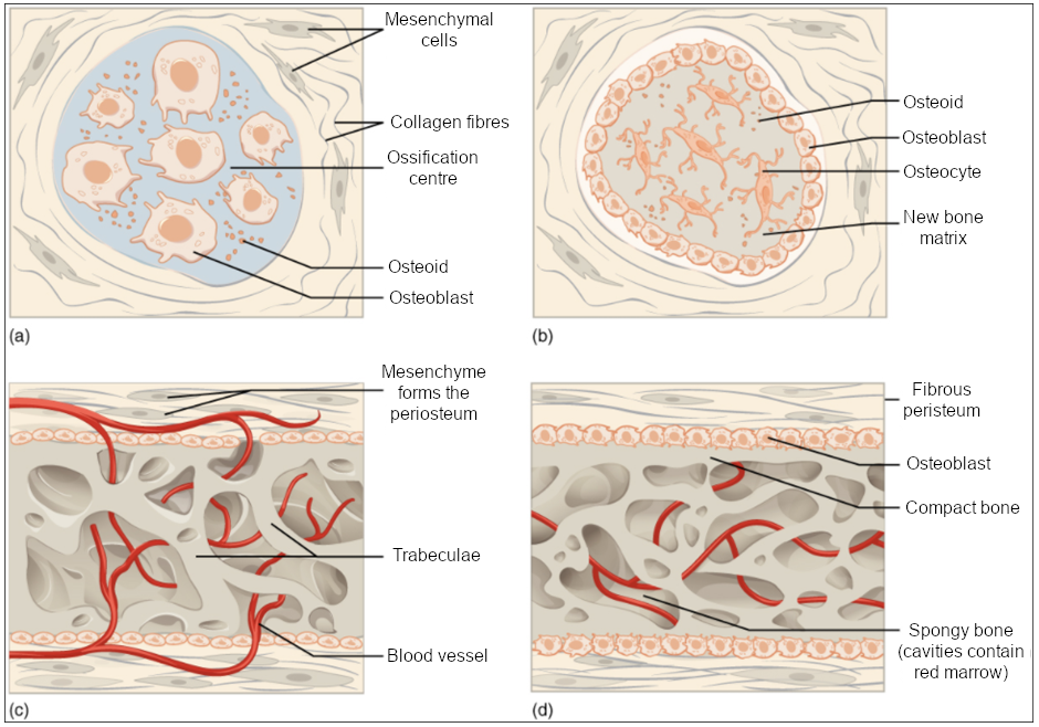
To summarize the key steps:
- Ossification Center Forms: Mesenchymal cells cluster together and differentiate into osteoblasts. This cluster is called an ossification center.
- Osteoid Secretion and Calcification: The osteoblasts secrete osteoid (the uncalcified bone matrix we discussed in the last lesson). Within a few days, calcium salts are deposited, and the matrix hardens. Osteoblasts that get trapped in the matrix mature into osteocytes.
- Woven Bone and Periosteum Formation: The osteoid is laid down around embryonic blood vessels, forming a network of trabeculae. This initial, immature bone is called woven bone. The surrounding mesenchyme condenses to form the periosteum.
- Lamellar Bone Replaces Woven Bone: The woven bone at the surface is remodeled into a layer of compact bone, with spongy bone remaining in the center. The blood vessels within the spongy bone become red marrow.
A key clinical connection for this process is the fontanelles, or "soft spots," on a newborn's skull. These are areas of fibrous membrane that have not yet undergone intramembranous ossification. Their flexibility allows the skull to deform during birth and accommodate the rapid brain growth that occurs in infancy.
Endochondral Ossification: Replacing a Cartilage Model
The term "endochondral" means "inside cartilage." This process is used to form most of the bones in the body, particularly long bones. It is more complex because it uses a hyaline cartilage model as a template, which is then systematically broken down and replaced by bone.
The key thing to remember is that cartilage does not transform into bone. It is a scaffold that is replaced.
This process is more involved, so we'll turn to a detailed video explanation.
Ossification | Bone Formation | Histogenesis of Bone | Bone Histology | Embryology of the Skeleton
The "Ossification" video from Byte Size Med provides a clear, animated walkthrough of endochondral ossification.
First, watch the brief introduction from this section on endochondral ossification for an overview. Then, watch the detailed explanation of the process from this segment. Pay close attention to the concepts of the primary and secondary ossification centers and the epiphyseal growth plate.
Let's break down the major events you saw in the video:
- Cartilage Model Formation: Mesenchymal cells differentiate into chondroblasts, which produce a hyaline cartilage model in the shape of the future bone.
- Bone Collar and Primary Ossification Center: The perichondrium around the diaphysis (shaft) becomes the periosteum. Osteoblasts secrete a thin layer of bone called the bone collar. Meanwhile, chondrocytes in the center hypertrophy (enlarge) and their matrix calcifies, cutting them off from nutrients and causing them to die.
- Vascular Invasion: Blood vessels invade the deteriorating cartilage, bringing osteoblasts and osteoclasts with them. The osteoblasts begin depositing bone matrix on the remnants of calcified cartilage, forming the primary ossification center in the diaphysis.
- Medullary Cavity Formation: As the primary ossification center grows towards the ends of the bone, osteoclasts break down the newly formed spongy bone in the very center, creating the medullary cavity.
- Secondary Ossification Centers: After birth, a similar process occurs in the epiphyses (the ends of the bone), where secondary ossification centers develop.
- Epiphyseal Plate and Articular Cartilage: The replacement process leaves cartilage in only two places:
- Articular cartilage on the outer surface of the epiphyses, which cushions the joint.
- The epiphyseal plate (or growth plate), a layer of hyaline cartilage between the diaphysis and epiphysis that allows the bone to grow in length during childhood and adolescence.
Comparing the Two Processes
Now that we've seen both pathways, let's put them side-by-side to solidify the comparison. The following reading activity asks a question that directly addresses our learning outcome.
6.4 Bone Formation and Development - Oregon State University
This question from the Chapter Review of the OpenStax resource provides a perfect summary comparison.
Read the question "In what ways do intramembranous and endochondral ossification differ?" and then click to reveal and study the detailed answer provided.
To summarize the comparison in a simple table for your exam review:
| Feature | Intramembranous Ossification | Endochondral Ossification |
|---|---|---|
| Starting Material | Fibrous mesenchymal membrane | Hyaline cartilage model |
| Process | Direct (Mesenchyme → Bone) | Indirect (Mesenchyme → Cartilage → Bone) |
| Ossification Centers | Multiple, form at the same time | Primary (1) and Secondary (2+) |
| Bones Formed | Flat bones of skull, mandible, clavicles | Most bones, especially long bones |
| Key Feature | Forms fontanelles in the skull | Involves an epiphyseal growth plate |
Conclusion
In this lesson, we have compared the two fundamental methods of bone development. You now know how different parts of your skeleton came to be.
Key Takeaways:
- Intramembranous ossification is a direct process where bone forms within a fibrous membrane. It is responsible for creating flat bones like those in the skull.
- Endochondral ossification is an indirect process where bone replaces a pre-existing hyaline cartilage model. It is responsible for forming long bones and most other bones in the skeleton.
- The key difference is the starting template: a mesenchymal membrane versus a cartilage model.
- Both processes ultimately produce the same mature bone tissue, composed of compact and spongy bone.
We've now covered how bone is built from the ground up. But bone is not a static structure; it's constantly being broken down and rebuilt throughout your life. In our next lesson, we will explore this dynamic process of bone remodeling and see how the balance between osteoblast and osteoclast activity maintains skeletal health and regulates calcium levels in the body.