Welcome back! In our last session, we explored the surfaces of bones, learning the language of markings that reveal a bone's function and its interactions with muscles and nerves. We moved from a macroscopic view of bone shapes to the specific features on their exteriors.
Today, we take the next logical step and zoom in even further, transitioning from gross anatomy to histology. We will examine the microscopic architecture of compact bone, the incredibly dense and strong tissue that forms the outer shell of our bones. By the end of this lesson, you will be able to describe the key components that give compact bone its remarkable properties: the osteon, lamellae, lacunae, and canaliculi. Understanding this intricate structure is fundamental to appreciating how bones can be both rigid supports and dynamic, living organs.
The Architecture of Strength: Compact Bone
Compact bone, also known as cortical bone, accounts for about 80% of the bone mass in your skeleton. It's what you picture when you think of a typical smooth, white bone. This tissue isn't just a solid, inert block; it's a highly organized and vital material, engineered for strength and resilience. Its fundamental structural and functional unit is the osteon.
To get a clear picture of how compact bone is built, we'll watch a detailed video that breaks down its microscopic organization.
Structure of Bone | Lamellar Bone | Compact and Cancellous Bone | Bone Histology
This video from Byte Size Med provides an excellent, detailed walkthrough of the microscopic structure of mature compact bone.
Please watch the segment from the beginning of the discussion on lamellar bone. Pay close attention to the descriptions of lamellar bone, the Haversian (central) canal, concentric lamellae, osteocytes in their lacunae, and the canaliculi. The video explains how these components assemble to form an osteon, or Haversian system.
The video you just watched introduced the core building block of compact bone: the osteon. Let's break down its components piece by piece, using some diagrams to help visualize their relationships.
Components of the Osteon
An osteon is essentially a microscopic, cylindrical pillar within the bone. Think of compact bone as a dense bundle of these pillars running parallel to the bone's long axis, making it incredibly strong at resisting compression and bending.

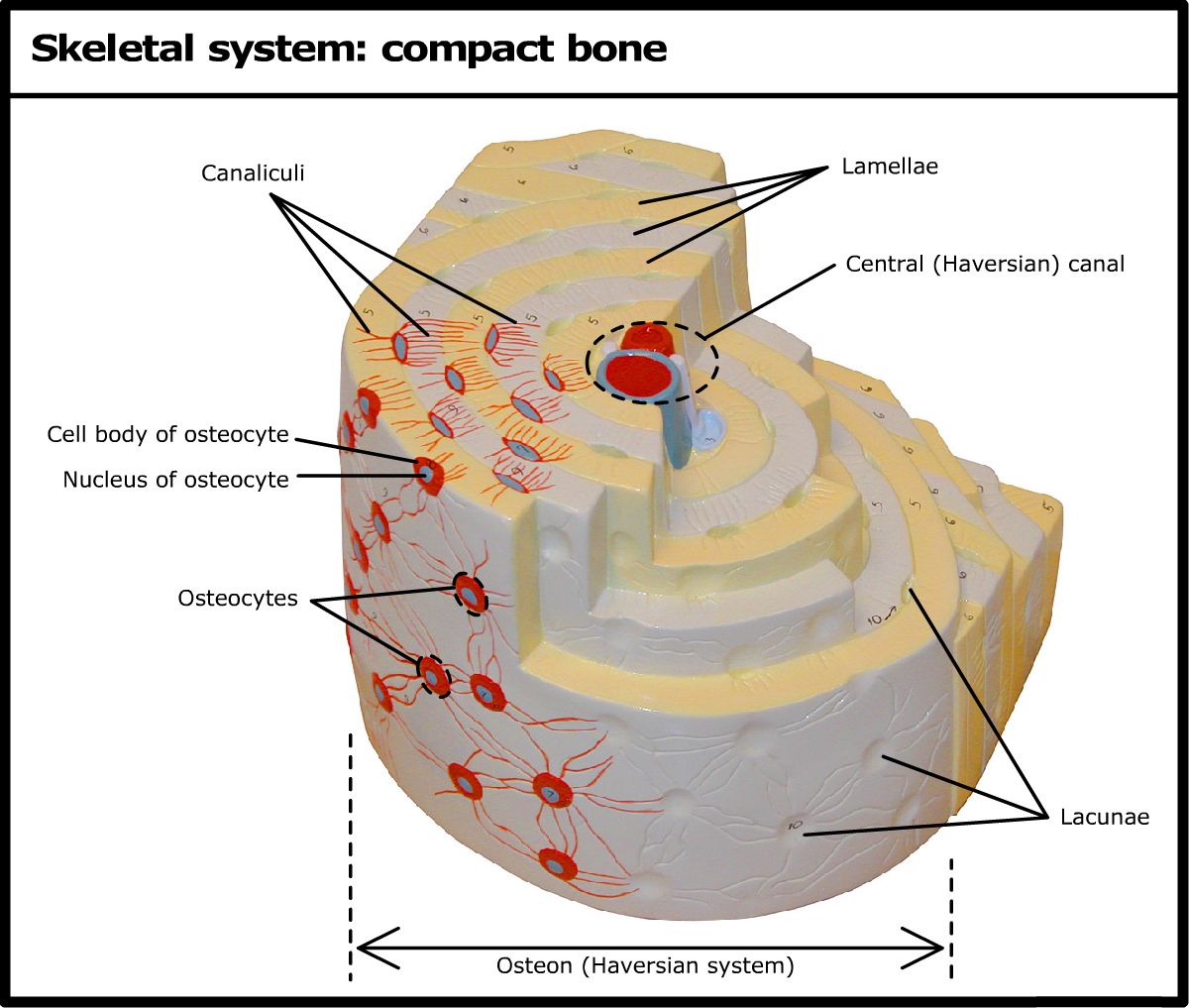
Let's examine the parts labeled in this diagram:
-
Central Canal (Haversian Canal): At the very center of each osteon is a hollow channel called the central canal. This is not empty space; it serves as a vital conduit for the bone's blood vessels and nerve fibers. The presence of this vascular network is what makes bone a living tissue, capable of healing and remodeling.
-
Lamellae: These are the concentric rings of bone matrix that surround the central canal, resembling the rings of a tree trunk. Each lamella is a layer of calcified extracellular matrix. This matrix is composed mainly of collagen fibers and inorganic calcium salts (hydroxyapatite), a combination that gives bone both flexibility (from collagen) and hardness (from calcium).
-
Lacunae and Osteocytes: Nestled in small cavities between the lamellae are the lacunae (singular: lacuna, meaning "little lake"). Inside each lacuna resides a mature bone cell, an osteocyte. These cells are responsible for maintaining the bone matrix and sensing mechanical stress. We will explore their full function in a future lesson.
-
Canaliculi: Radiating outward from the central canal and connecting the lacunae are tiny, hair-like channels called canaliculi (singular: canaliculus, meaning "little canal"). Because the calcified bone matrix is too dense for nutrients to diffuse through, the canaliculi form a crucial transport network. They allow osteocytes to connect with each other and with the blood supply in the central canal, receiving nutrients and removing waste.
A Masterpiece of Engineering
The arrangement of these components is not random. It's a highly efficient system that maximizes strength while maintaining the living tissue. A particularly brilliant feature is the organization of collagen fibers within the lamellae.
To learn more about this, let's turn to a concise textual resource that explains this structural detail.
Microscopic Anatomy of Bones - The Osteon - Pearson
This text from Pearson's A&P channel explains the composition of lamellae and how the arrangement of collagen fibers contributes to bone's torsional strength.
Please read the section under the heading Structure of Lamellae. Focus on how the alternating orientation of collagen fibers in adjacent lamellae helps the bone resist twisting forces.
As you just read, the collagen fibers in one lamella run in a single direction, but the fibers in the adjacent lamella run in the opposite direction. This alternating, helical pattern makes the osteon exceptionally strong at resisting torsion, or twisting forces—a common stress on long bones.
The Bigger Picture: Connecting the Osteons
Finally, it's important to understand how these individual osteons are integrated into the larger structure of compact bone. The central canals run longitudinally down the bone, but they are not isolated. They are connected by transverse channels called perforating canals (or Volkmann's canals). These canals run perpendicular to the central canals and link the blood and nerve supplies of the periosteum (the outer membrane of the bone) to the central canals and the medullary cavity within.
To consolidate everything we've covered, please read this summary.
Microscopic Anatomy of Bones - The Osteon - Pearson
This text provides a final, clear summary of all the components of the osteon and their functions.
First, read the main description of the osteon. Then, review the explanations for Canaliculi and Perforating canals in the Q&A section at the bottom.
Conclusion
In this lesson, we have magnified our view to the microscopic level to understand the intricate and elegant structure of compact bone. We've seen that it's far from being a simple, solid material. Instead, it is a dynamic, living tissue built from highly organized functional units.
Key Takeaways:
- The primary structural unit of compact bone is the osteon (or Haversian system).
- Each osteon consists of a central Haversian canal containing blood vessels and nerves, surrounded by concentric layers of bone matrix called lamellae.
- Mature bone cells, or osteocytes, reside in small chambers called lacunae situated between the lamellae.
- A network of tiny channels, the canaliculi, connects the lacunae, allowing for nutrient transport and communication between osteocytes.
- Perforating canals connect adjacent osteons, integrating the entire bone into a single vascular and neural network.
We have now explored the dense outer layer of bone. However, the inside of many bones looks very different. In our next lesson, we will investigate the structure of spongy (cancellous) bone and compare its form and function to the compact bone we studied today.