Welcome back! In our last lesson, we took a deep dive into the molecular engine of muscle contraction: the cross-bridge cycle. We saw how ATP, calcium, troponin, and tropomyosin work together in a four-step process that allows myosin heads to pull on actin filaments.
Today, we'll zoom out from that single molecular interaction to understand how the coordinated action of millions of these "power strokes" leads to the shortening of a muscle fiber. Your learning outcome for this lesson is to explain the sliding filament mechanism of muscle contraction as the result of sarcomere shortening. We will see how the components of the sarcomere change during contraction and connect this large-scale movement back to the molecular cycle you've already mastered.
The Central Idea: Filaments Slide, They Don't Shrink
Let's begin with the foundational concept of this mechanism. When a muscle contracts, its individual protein filaments—actin and myosin—do not get shorter. Instead, the thin (actin) filaments slide past the thick (myosin) filaments, increasing their overlap. This sliding action pulls the ends of the sarcomere closer together, causing the entire unit to shorten. Since muscle fibers are essentially long chains of sarcomeres connected end-to-end, the shortening of all sarcomeres results in the contraction of the entire muscle.
The following video provides an excellent, straightforward explanation of this core principle.
Muscle Tissues and Sliding Filament Model
This clip from the "Muscle Tissues and Sliding Filament Model" video by Amoeba Sisters clearly illustrates the central concept of the sliding filament mechanism.
Please watch from the main concept. Pay close attention to the statement that the filaments themselves do not shorten, but instead slide past each other to shorten the sarcomere.
To help visualize this, let's use a simple analogy.
The Sliding Filament Theory of Muscle Contraction
This text from Nature Education offers a helpful analogy to make the sliding filament concept more concrete.
Read the section titled An Analogy for Sliding Filaments. As you read, picture yourself as the myosin filament, the ropes as the actin filaments, and the bookcases as the Z discs.
This analogy beautifully captures the essence of the mechanism: the central element (you/myosin) stays in place while pulling the outer elements (bookcases/Z discs) inward via the connecting ropes (actin).
A Closer Look: What Happens to the Sarcomere Bands?
Now, let's apply this sliding concept to the specific bands and zones of the sarcomere that you've learned about. During contraction, the visible structure of the sarcomere changes in a predictable way.
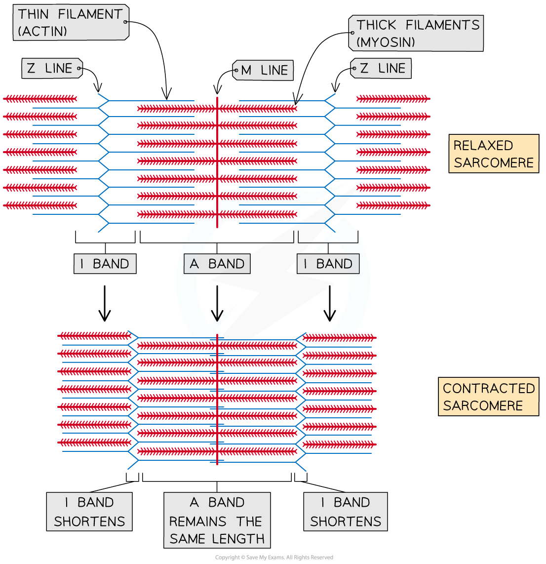
Here is a breakdown of what happens to each part of the sarcomere during contraction, as shown in the diagram above:
- Z discs: These are the boundaries of the sarcomere. As the thin filaments are pulled inward, the Z discs move closer together, causing the entire sarcomere to shorten.
- I Band: This is the region containing only thin filaments. As the thin filaments slide over the thick filaments, the I band shortens significantly.
- H Zone: This is the central region of the A band containing only thick filaments. As the thin filaments are pulled toward the M line, they invade the H zone, causing it to narrow and eventually disappear in a maximal contraction.
- A Band: This band represents the entire length of the thick myosin filaments. Because the thick filaments themselves do not change length, the A band's length remains constant throughout the contraction.
The observation that the A band remains constant while the I band shortens was the key piece of evidence that led scientists to propose the sliding filament theory in the 1950s.
To solidify these details, the following text provides a concise summary of the sarcomere zones and what happens to them.
Ultrastructure of Muscle - Skeletal - Sliding Filament
This article from TeachMeAnatomy clearly summarizes the changes within the sarcomere during contraction.
Please read the section under the heading Sliding Filament Model. Focus on the final paragraph of this section, which explicitly states which bands shorten (H zone and I band) and which stays the same (A band).
Linking the Cycle to the Slide
We can now connect the cross-bridge cycle (the "engine") to the sliding filament mechanism (the "movement"). The four steps of the cross-bridge cycle—attachment, power stroke, detachment, and reactivation—are what drive the sliding motion.
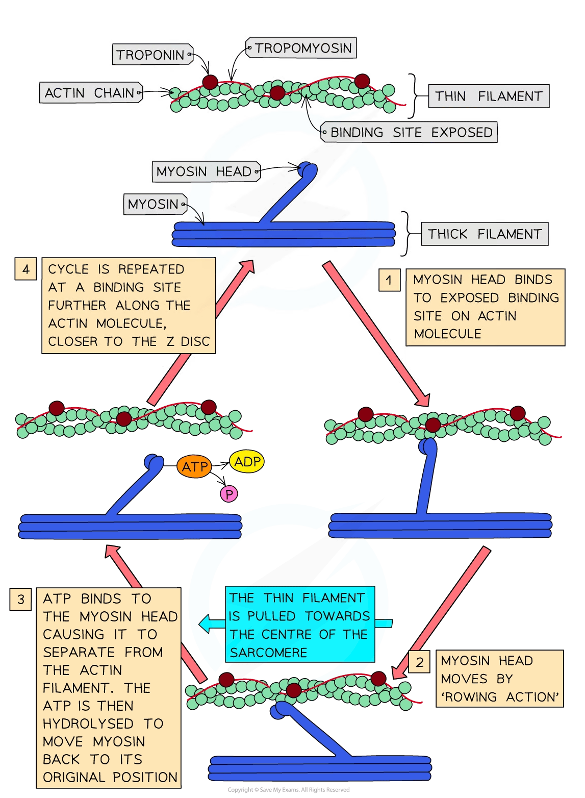
Imagine thousands of myosin heads on a single thick filament, all cycling at slightly different times. Each power stroke contributes a tiny "tug" on the actin filament. The sum of all these asynchronous tugs results in a smooth, continuous sliding of the thin filament past the thick filament. It's like a team of rowers paddling a boat; not all oars are in the water at once, but their collective, repeated efforts produce continuous forward motion.
This short video clip nicely summarizes this final connection.
Muscles, Part 1 - Muscle Cells: Crash Course Anatomy & Physiology #21
This segment from Crash Course's "Muscles, Part 1" directly links the power stroke to the sliding motion.
Watch from this description of the pull. The narrator explains that when the myosin head changes shape, it pulls on the actin strand, shrinking the sarcomere. This is the "sliding" part of the model.
The process continues as long as calcium is present to keep the actin binding sites exposed and ATP is available to power the myosin heads. When the nerve signal ceases, calcium is pumped back into the sarcoplasmic reticulum, troponin and tropomyosin return to their blocking positions, and the muscle fiber relaxes as the sarcomeres return to their original length.
Conclusion
In this lesson, we have bridged the gap between molecular action and muscular contraction. You can now explain how the repetitive binding and pulling of the cross-bridge cycle drives the sliding of actin and myosin filaments, resulting in the shortening of the sarcomere. This is the fundamental mechanism that enables every voluntary movement, from lifting a book to running a marathon.
Key Takeaways:
- The sliding filament mechanism describes muscle contraction as the sliding of thin (actin) filaments past thick (myosin) filaments.
- The filaments themselves do not shorten; the sarcomere shortens because the overlap between the filaments increases.
- During contraction, the I bands and H zones shorten, while the A band remains constant in length. The Z discs are pulled closer together.
- The cross-bridge cycle provides the mechanical force for this sliding. Each "power stroke" is a small pull that, when repeated by millions of myosin heads, creates the overall contraction.
Now that you understand the fundamental mechanism of how a single muscle fiber contracts, the next logical step in understanding muscle physiology is to explore how the nervous system controls the strength and duration of a contraction in a whole muscle. This involves concepts like motor units, recruitment, and how the frequency of stimulation affects muscle tension.