Welcome to your next lesson. In our last session, we finished our tour of the skull by identifying the crucial foramina that serve as passages for nerves and blood vessels. We ended at the foramen magnum, the large opening where the brainstem connects to the spinal cord. Today, we will follow that connection downward to explore the structure that protects the spinal cord: the vertebral column.
Your learning goal for this lesson is to identify the five regions and four curves of the vertebral column. This is the foundational knowledge for understanding posture, movement, and common clinical issues like back pain and spinal injuries, which you will frequently encounter in your nursing career.
The Regions of the Vertebral Column
The vertebral column, or spine, is not a single rigid rod but a flexible column made of 24 individual vertebrae, plus the fused bones of the sacrum and coccyx. To make sense of it, anatomists divide it into five distinct regions.
Let's begin with a clear overview of these regions.

To learn the names of these regions and the number of vertebrae in each, let's watch a short, helpful video. The presenter offers some useful mnemonics to aid in memorization, which will be valuable for your exam preparation.
Vertebral Column Anatomy: Bones, Regions, Curvatures (Kyphotic, Lordotic)
This video, "Vertebral Column Anatomy" from RegisteredNurseRN, provides a clear and concise breakdown of the five spinal regions.
Please watch the section covering the five main regions. Pay close attention to the name, abbreviation, and number of vertebrae for each region.
As a quick summary of what you just watched, here are the five regions of the vertebral column from superior to inferior:
- Cervical Region (C1–C7): Comprises the 7 vertebrae of the neck.
- Thoracic Region (T1–T12): Comprises the 12 vertebrae of the upper and mid-back. Each of these articulates with a pair of ribs.
- Lumbar Region (L1–L5): Comprises the 5 large vertebrae of the lower back, which bear the majority of the body's weight.
- Sacrum (S1–S5): A single bone in adults, formed from the fusion of 5 vertebrae. It connects the spine to the hip bones.
- Coccyx (Co1–Co4): The "tailbone," also a single bone in adults, formed from the fusion of 3 to 5 small vertebrae.
The Curvatures of the Vertebral Column
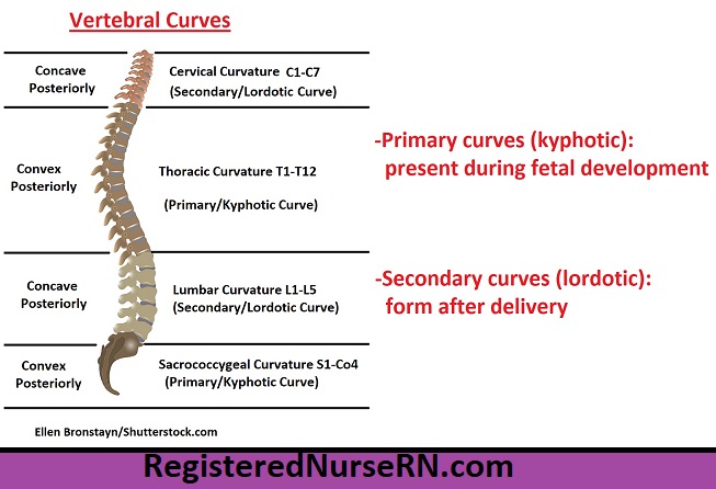
Looking at the spine from the side, it's clear it is not a straight line. It has four distinct curves that give it an S-shape. These curves are not a defect; they are crucial for providing strength, flexibility, and the ability to absorb shock and distribute body weight effectively.
The four curves are classified into two types based on when they develop.
- Primary Curves: These are present at birth and retain the original C-shape of the fetal spine. They are also called kyphotic curves.
- Secondary Curves: These develop after birth as a child learns to hold up their head, sit, and walk. They are also called lordotic curves.
The following image provides an excellent visual summary of these curves.
Now, let's return to the video from RegisteredNurseRN to hear a guided explanation of these curves.
Vertebral Column Anatomy: Bones, Regions, Curvatures (Kyphotic, Lordotic)
This segment of the video explains the development and names of the four spinal curvatures.
Please watch the clip focusing on the curves of the spine. Note which curves are primary and which are secondary, and the terms kyphotic and lordotic.
To deepen your understanding of why these curves form and their function, the following text provides a more detailed explanation.
The Vertebral Column | Anatomy and Physiology I
This reading from Lumen Learning explains the functional importance of the spinal curves and links their development to key infant milestones.
Please read the section titled "Curvatures of the Vertebral Column", focusing on the description of primary and secondary curves. Then, continue reading to learn how the cervical and lumbar curves develop as a child grows.
To bring all this information together, here is a summary of the four curves:
| Curve Name | Region | Type | Curvature | Develops |
|---|---|---|---|---|
| Cervical | C1–C7 | Secondary | Concave posteriorly | When an infant lifts its head |
| Thoracic | T1–T12 | Primary | Convex posteriorly | During fetal development |
| Lumbar | L1–L5 | Secondary | Concave posteriorly | When a child begins to walk |
| Sacrococcygeal | Sacrum & Coccyx | Primary | Convex posteriorly | During fetal development |
An excessive exaggeration of these natural curves can lead to clinical conditions. An abnormal posterior curve in the thoracic region is called kyphosis (hunchback), while an excessive anterior curve in the lumbar region is known as lordosis (swayback).
Conclusion
In this lesson, you have learned the fundamental organization of the vertebral column. We moved down from the skull to identify the five regions that make up the spine and the four curves that give it strength and resilience.
Key Takeaways:
- The vertebral column is divided into five regions: cervical (7 vertebrae), thoracic (12), lumbar (5), the sacrum (5 fused), and the coccyx (3-5 fused).
- The adult spine has four curvatures that help with shock absorption and weight-bearing.
- The thoracic and sacrococcygeal curves are primary curves (kyphotic), present from fetal development.
- The cervical and lumbar curves are secondary curves (lordotic), which develop after birth as a child gains motor control.
Now that you have this broad overview of the vertebral column's structure, our next lesson will zoom in on the vertebrae themselves. We will learn to distinguish between cervical, thoracic, and lumbar vertebrae based on their key structural features.