Welcome to the first lesson of your journey into the science of breathwork. To achieve your goal of building a personalized system of breathing techniques, we must first establish a solid foundation in the underlying physiology. This initial module focuses on the fundamental mechanics of the respiratory system.
In this lesson, you will learn to describe the mechanics of inhalation and exhalation. We will explore the precise roles of the key muscles—the diaphragm and the intercostals—and distinguish between the passive nature of quiet exhalation and the active muscular effort required for forceful breathing. Understanding this physical machinery is the first critical step toward deconstructing any breathwork practice, from ancient Pranayama to modern clinical techniques.
The Engine of Breath: Volume, Pressure, and Boyle's Law
At its core, breathing is a physical process governed by a simple principle: air flows from an area of higher pressure to an area of lower pressure. The elegant function of your respiratory system is to continuously create pressure gradients between the atmosphere and your lungs.
The physical law that dictates this relationship is Boyle's Law, which states that for a fixed amount of gas at a constant temperature, pressure and volume are inversely proportional.
This means that if you increase the volume of a container, the pressure of the gas inside it decreases, and vice versa. Your thoracic cavity (your chest) is that container. By changing its volume, you change the pressure within your lungs, causing air to either rush in or be pushed out.
To see a clear overview of this principle and the key players involved, please watch the following introductory video.
This video from Dr Matt & Dr Mike, "Basic Breathing Mechanics," provides an excellent introduction to the respiratory cycle and the application of Boyle's Law.
Watch the segment from the beginning until the end of the explanation on inspiration. Pay close attention to the syringe analogy, as it's a perfect illustration of how increasing volume creates negative pressure to draw air in.
The Mechanics of Inhalation (Inspiration)
Quiet, restful inhalation is an active process driven by the contraction of two main muscle groups.
-
The Diaphragm: This large, dome-shaped muscle sits at the base of your thoracic cavity, separating it from the abdominal cavity. At rest (during exhalation), it is relaxed and curved upwards. When it contracts, it flattens and moves downwards, increasing the vertical dimension (height) of the thoracic cavity. This single action is responsible for about 75% of the air movement in quiet breathing.
-
The External Intercostal Muscles: These are a series of muscles located between your ribs. When they contract, they pull your rib cage upwards and outwards. This movement increases the front-to-back (anteroposterior) and side-to-side (lateral) dimensions of your thoracic cavity.
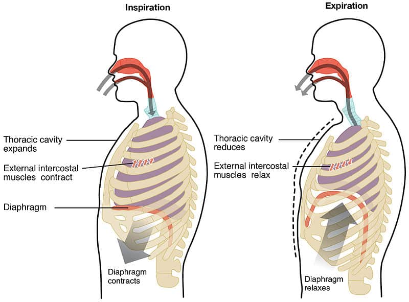
The combined action of the diaphragm and external intercostals expands the volume of the thoracic cavity in all three dimensions. As Boyle's law dictates, this increase in volume causes the pressure inside the lungs (intrapulmonary pressure) to drop slightly below atmospheric pressure, creating a pressure gradient that draws air into the lungs until the pressures equalize.
For a more detailed mechanical and neurological breakdown of inspiration, the following video provides an exceptionally thorough explanation.
Respiratory | Mechanics of Breathing: Inspiration | Part 2
The video "Respiratory | Mechanics of Breathing: Inspiration" by Ninja Nerd offers a deep dive into the anatomy and physiology of inhalation. It is more detailed, which will be valuable for your analytical approach.
First, watch the section on muscles and nerves to understand the anatomical locations and the specific nerves (phrenic and intercostal) that control the diaphragm and external intercostals. Next, focus on the explanation of the "bucket handle" and "old school water pump" actions from the start of this section. This will give you a three-dimensional mental model of how the rib cage expands. Finally, observe how these movements translate into pressure changes from intrapleural to intrapulmonary pressure, and how the resulting pressure gradient drives airflow from high to low pressure.
The Mechanics of Exhalation (Expiration)
How you exhale depends on your level of activity.
Passive Exhalation
During quiet breathing, exhalation is a passive process. It does not require active muscle contraction. Instead, it relies on two factors:
- Muscle Relaxation: The diaphragm and external intercostal muscles simply relax, returning to their resting positions.
- Elastic Recoil: Lung tissue is rich in elastic fibers. After being stretched during inhalation, the lungs and the chest wall naturally recoil to their smaller, resting size.
This decrease in thoracic volume increases the intrapulmonary pressure to slightly above atmospheric pressure, pushing air out of the lungs. The video from Dr. Matt & Dr. Mike that you watched earlier explains this process well.
Active (Forceful) Exhalation
Forceful exhalation, such as during exercise, speaking, or many breathwork techniques (e.g., Kapalabhati, Breath of Fire), is an active process that recruits additional muscles:
- The Abdominal Muscles: This group includes the rectus abdominis, transverse abdominis, and the internal and external obliques. When they contract, they increase pressure within the abdominal cavity, pushing the diaphragm upwards forcefully and rapidly.
- The Internal Intercostal Muscles: Located beneath the external intercostals, these muscles have the opposite action. When they contract, they pull the rib cage down and inward, actively reducing the volume of the thoracic cavity.
You can feel this process by placing a hand on your abdomen and coughing. The sharp contraction you feel is your abdominal wall engaging to expel air forcefully.
To complete the picture, return to the "Basic Breathing Mechanics" video.
Watch the final segment on passive and active expiration. This will solidify your understanding of how the internal intercostals and abdominal muscles contribute to forceful exhalation.
An Evidence-Based View: Quantifying Muscle Roles
Given your expertise in analyzing clinical data, you will appreciate that these muscle functions are not just theoretical. They are quantified in research through methods like surface electromyography (sEMG), which measures the electrical activity produced by muscle contractions.
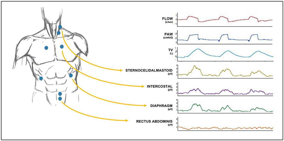
The following research paper provides an excellent example of how scientists study the relative contributions and fatigue of the primary inspiratory muscles. You don't need to read the entire paper, but focusing on a few key sections will demonstrate how the roles of these muscles are experimentally validated.
Assessment of Diaphragm and External Intercostals Fatigue ... - MDPI
This study from MDPI's Sensors journal investigates inspiratory muscle fatigue after maximal voluntary ventilation (MVV), a task that pushes the respiratory system to its limits.
First, read the Abstract to get a high-level summary of the study's design and conclusions. Next, review section 2.5 on sEMG recordings to understand the methodology for data collection, including electrode placement as shown in their Figure 1. Then, examine Table 2 under the Results section, which presents the core data. Focus on the "Diaphragm" and "External intercostals" rows. Notice the changes in RMS (μV), a measure of electrical amplitude, before (Pretest) and immediately after (Posttest) the MVV maneuvers. You'll see a statistically significant decrease for the diaphragm but not for the external intercostals. Finally, read the second paragraph of the Discussion, starting from "EMG RMS amplitude...". The authors interpret the data, suggesting that because the diaphragm shows significant fatigue while the intercostals do not, the diaphragm is the "principal pressure generator" during these high-demand maneuvers.
This study provides direct, quantitative evidence for the diaphragm's primary role in breathing, a central concept we will build upon throughout this course.
Another research method, dynamic magnetic resonance imaging (D-MRI), allows us to visualize and measure the physical movement of the chest wall and diaphragm. A study in the Journal of Clinical Medicine found that during both quiet and deep breathing, chest wall motion is most prominent in the anterior-posterior direction, confirming the "pump handle" action. It also showed that the apex and posterior portions of the diaphragm move more than the anterior portion, indicating its movement is complex and not entirely uniform. This complements the EMG data by showing the physical consequence of the electrical activation.
Conclusion
In this lesson, we have established the fundamental mechanical principles of breathing. Let's summarize the key takeaways:
- Breathing is driven by pressure gradients, which are created by changing the volume of the thoracic cavity according to Boyle's Law.
- Quiet inhalation is an active process led by the downward contraction of the diaphragm and the upward-and-outward contraction of the external intercostal muscles.
- Quiet exhalation is a passive process that relies on the relaxation of these muscles and the elastic recoil of the lungs and chest wall.
- Forceful exhalation is an active process that recruits the internal intercostals and abdominal muscles to rapidly and forcefully decrease thoracic volume.
- Experimental evidence from EMG and MRI studies confirms that the diaphragm is the primary muscle of inspiration, responsible for the majority of air movement.
With this mechanical framework in place, you are now equipped to understand how different breathing techniques manipulate the body.
In our next lesson, we will zoom in from the level of gross anatomy to the microscopic level of the alveoli. We will explore gas exchange, explaining how oxygen moves from the air you've just inhaled into your bloodstream, and how carbon dioxide makes the reverse journey.