Hello. Last lesson explained why a keloid is more than an ordinary scar: scar-forming activity produces excess tissue that may spread beyond the injury. Another kind of raised scar, the hypertrophic scar, can also contain excess scar tissue. This lesson focuses on how to tell the two patterns apart in a short history. The key is not simply how raised a mark looks, but where it grows, when it appears, and what it does over time.
Start with the original wound
Imagine drawing a boundary around the skin that was injured. A hypertrophic scar becomes thick or raised within that boundary. A keloid grows beyond it, into surrounding skin that was not part of the original wound. Both can feel firm, and both can itch or cause discomfort. Height, firmness, and itching alone therefore do not settle the distinction.
This short video makes the boundary difference visible:
Hypertrophic Scar vs. Keloid: What’s the Difference?
Watch “Hypertrophic Scar vs. Keloid: What’s the Difference?” by Sam Lam for a visual explanation of why a raised scar is not necessarily a keloid.
Watch the border comparison. Focus on whether the raised tissue remains inside the initial injury or occupies a wider area. Treat the large size examples as illustrations, not as a minimum size required for a keloid.
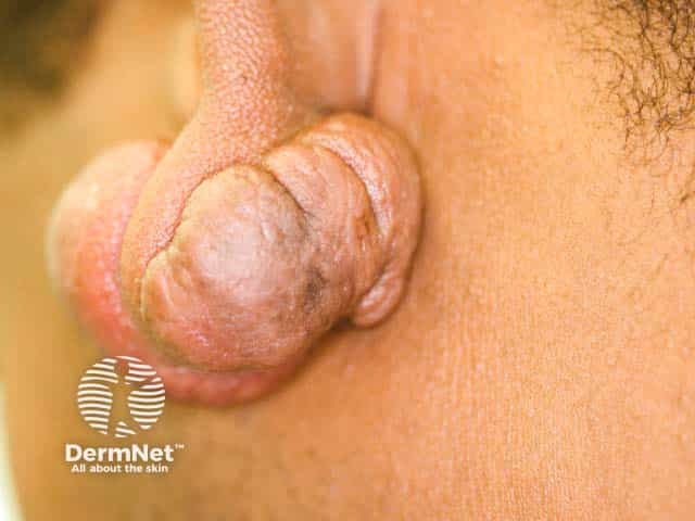
An earlobe piercing shows why this distinction can be harder to apply than it sounds. The original puncture is small, but by the time a bump is noticed, the initial wound may no longer be visible. The photograph below shows an earlobe keloid; the image alone cannot tell you when it began or reconstruct the precise boundary of the piercing injury.
Add time to the picture
The previous lesson described scar repair as a process that changes over time. That history helps distinguish these two raised scars. A hypertrophic scar usually appears within weeks of an injury. It may become more prominent for a while, then soften or flatten over subsequent months. A keloid may first become noticeable months or even years later and is more likely to persist or continue enlarging.
Read DermNet’s comparison with those three clues—boundary, onset, and later course—in mind:
Hypertrophic Scars and Keloids: A Complete Overview — DermNet
Read DermNet’s overview to see how clinicians combine the scar’s appearance with its history, rather than relying on raised appearance alone.
In “What are keloids and hypertrophic scars?”, read the definitions; pay particular attention to the original wound margin. In “What are the clinical features of keloids and hypertrophic scars?”, begin at “Keloids are usually:” and read both feature lists, comparing onset and growth as well as appearance. Finally, in “How are keloids and hypertrophic scars diagnosed?”, read the diagnostic discussion. Stop before the paragraphs about microscopic findings.
These are typical patterns, not a stopwatch test. A hypertrophic scar does not have to disappear within a set number of months; some remain raised. A keloid need not grow continuously at a steady rate. Early appearance does not rule out a keloid, and persistence by itself does not prove one. Growth beyond the original injury is the strongest distinguishing clue, interpreted alongside timing and later change.
Read a short history, not just a snapshot
Consider a cut that heals, then develops a thick red ridge along the cut a few weeks later. Six months on, the ridge is still visible but softer, and it has never spread beyond the cut. That history is more consistent with a hypertrophic scar: early onset, confinement to the wound, and some regression all point in the same direction.
Now consider a piercing that seems to heal, followed months later by a firm growth that becomes wider than the original injured area and remains prominent. That history is more consistent with a keloid. The decisive detail is not that the growth is large or raised; it is that scar tissue has extended past the initial injury.
A less complete history might say only, “There is a raised bump beside a recent piercing.” It gives neither a reliable original boundary nor a course over time. Calling it a keloid—or a hypertrophic scar—on that description would be premature. Other causes of piercing bumps will be covered later in the course.
Clinicians use the history and examination together, and the distinction can still be difficult. If a lump’s origin or behavior is unclear, a clinician can assess it rather than relying on a photograph or a label assigned online.
Conclusion
Both keloids and hypertrophic scars are raised forms of excess scarring. To distinguish them in a case, first compare the growth with the original wound boundary, then use onset and persistence as supporting evidence. Hypertrophic scars generally arise sooner, stay within the injured area, and are more likely to soften; keloids extend beyond the injury and are more likely to persist. None of those tendencies makes raised appearance alone a diagnosis.
Next, the course turns to why some piercing or tattoo injuries carry more keloid risk than others—and why risk factors cannot predict an individual scar with certainty.