Hello. The previous lesson focused on when symptoms after exercise warrant urgent assessment. The next step is understanding what clinicians look for once that assessment begins. Creatine kinase (CK), creatinine, and electrolytes answer different questions: how much muscle has been injured, whether kidney function may be affected, and whether changes in the blood could cause immediate harm. None tells the whole story alone.
CK: evidence of muscle injury
When muscle cells break down, they release CK, an enzyme abundant in muscle, into the bloodstream. A substantially raised blood CK level, interpreted alongside recent exertion and symptoms, is the principal laboratory evidence used to identify rhabdomyolysis. But CK measures muscle injury; it does not directly measure kidney injury. A high result does not mean kidney failure has occurred, and an elevated CK after intense exercise does not, by itself, settle how serious the episode is.
Read the short section below for the distinction between CK and myoglobin. It also explains why clinicians interpret a CK result in context rather than relying on a universal number.
Exertional rhabdomyolysis: physiological response or manifestation of an underlying myopathy?
Read the “CK elevation” section of this clinical review to understand why CK is a more useful blood marker than myoglobin and why proposed CK cutoffs differ.
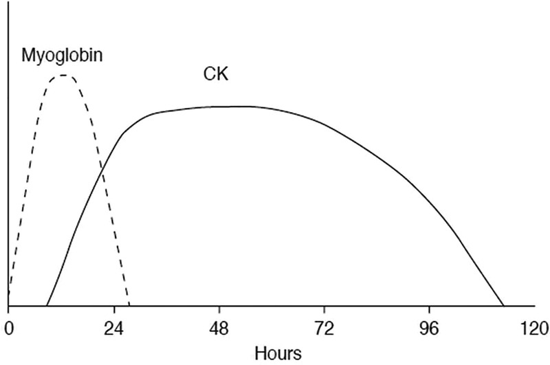
In “CK elevation,” read the marker comparison, including the Figure 2 caption. Then read the threshold discussion. Focus on the timing and interpretation; the exact numerical cutoffs are less important here.
Myoglobin—the muscle protein discussed earlier in the course—appears in blood earlier and clears relatively quickly. CK rises later and remains elevated longer, making it easier to detect muscle injury after the first hours have passed. The graph illustrates those relative patterns, not a schedule that every person follows.
A commonly used diagnostic approach considers CK several times above the laboratory’s upper limit of normal together with a compatible history and examination. Yet the degree of CK elevation can vary with the exercise and the person. Clinicians must also assess the consequences of muscle breakdown, not just confirm that it happened.
Creatinine: a different signal from the kidneys
Creatinine is not CK. It is a waste product measured in blood to help assess how well the kidneys are filtering. A rising creatinine level after suspected rhabdomyolysis can indicate developing kidney dysfunction. A previous result, if available, helps clinicians judge whether today’s value represents a change.
Creatinine also has limitations. A single value can be influenced by factors such as hydration and muscle-related differences between people, and kidney effects may not be apparent on the first test. Thus, an initially normal creatinine is encouraging but does not guarantee that kidney injury cannot develop. Conversely, an abnormal value needs clinical interpretation rather than automatic attribution to myoglobin.
The following table puts creatinine beside CK and the other tests clinicians use. Read only the indicated rows; the review contains additional specialist risk-assessment material that is not needed for this lesson.
Exertional rhabdomyolysis: physiological response or manifestation of an underlying myopathy?
Return to the clinical review for its comparison of muscle-injury, kidney-function, and electrolyte tests.
In “Step 2: what is the risk of developing ARF?”, read the opening paragraph for context. Then, in Table 4, “Assessment of risk of acute renal failure and other complications,” read the rows “Serum CK,” “Serum potassium,” “Serum sodium,” and “Renal function”: they run from CK through creatinine. Stop before the “Myoglobinuria” row. Notice that the tests describe different aspects of risk.
Electrolytes: changes that may need immediate attention
Electrolytes are substances such as potassium and sodium that help nerves, muscles, and the heart function. Injured muscle cells can release potassium and phosphate into the blood. A dangerously high potassium level can disturb heart rhythm, so it matters urgently even if a person’s CK has not yet peaked or their creatinine is still normal. Clinicians may check heart activity with an electrocardiogram when potassium is a concern.
Other results add context. Phosphate can rise, calcium can be low early in an episode, and sodium may be abnormal—for example, in someone with an exercise-related fluid imbalance. Electrolyte results help clinicians identify complications that CK alone would miss. They also require interpretation together: the aim is not to match each abnormal number to one cause without examining the person.
| Test | Its main contribution | What it cannot establish alone |
|---|---|---|
| CK | Evidence and course of muscle injury | Whether the kidneys are functioning normally |
| Creatinine | Evidence of possible change in kidney filtration | How much muscle has broken down |
| Electrolytes | Potentially dangerous blood-chemistry changes, especially potassium | Whether rhabdomyolysis is the cause of those changes |
Why the first set of results may not be the last
The time since exercise changes what a result means. Myoglobin can appear and clear early; CK may still be rising when someone first seeks care. Kidney-function and electrolyte results can also change as the episode develops. For these reasons, clinicians may repeat CK and a blood-chemistry panel rather than treating an early result as final.
The guideline below gives a concrete example: in a person assessed very soon after strenuous exercise, CK can initially be below a commonly used diagnostic threshold, and a clinician may arrange repeat testing around the following day. That is a clinical follow-up plan, not a reason to delay urgent assessment while waiting for a more revealing test.
Clinical Practice Guideline for the Management of ER
Read this guideline’s early-testing example to see why a first CK result may need to be revisited.
In section 4, “Evaluating Patients with Dark or ‘Cola-Colored’ Urine” (around printed p. 9), locate the discussion after the section’s opening paragraph about other possible causes of dark urine. Begin at the assessment discussion, ending with the example of repeating CK and a metabolic panel. Focus on the early timing and the reason to repeat blood tests, rather than the full list of alternative causes of dark urine.
Repeat results are useful because the direction of change matters. CK that is still rising suggests muscle injury may not yet have peaked. Creatinine that rises raises concern about kidney function, even if CK later begins to fall. A potassium level that becomes abnormal may require prompt action independently of either trend. Clinicians interpret these measurements alongside symptoms, examination, and urine output; there is no single repeat-testing interval appropriate for every situation.
Consider the contrast between two possible sets of findings. A person could have markedly elevated CK but currently normal creatinine and electrolytes: muscle injury is evident, while serious kidney or electrolyte complications are not evident at that moment. Another could have a less striking early CK result but dangerously high potassium: the lower CK provides no reassurance about that immediate risk. Both situations call for interpretation and follow-up appropriate to the whole clinical picture, not ranking by CK alone.
Key takeaways
CK helps identify and track muscle breakdown. Creatinine helps detect a possible effect on kidney function. Electrolyte tests reveal blood-chemistry complications, some of which can be urgent before kidney tests become abnormal. Because these markers change at different times, an early normal-looking result cannot always close the investigation; repeat measurements and clinical assessment show how the situation is evolving.
The next lesson turns to treatment: why clinicians use fluids to support the kidneys and why the amount and delivery of fluid must be guided by the person’s condition rather than by unsupervised excessive drinking.