Hello! Welcome back.
In our last lesson, we established a decision-making framework for when to perform a thyroid frozen section (FS), focusing on the indications and, crucially, the limitations. We concluded that the strongest indication is to confirm papillary thyroid carcinoma (PTC) in a nodule deemed "Suspicious for Malignancy" on FNA, while the greatest limitation is the inability to assess follicular-patterned lesions.
Today, we move from the why to the how. This lesson will focus on the practical interpretation of the slides themselves, addressing the learning outcome: Interpret key histologic features on frozen sections to distinguish benign from malignant processes. We will tackle the artifacts inherent to the technique and build a systematic approach to identifying the key features that allow for a confident intraoperative diagnosis—or a confident deferral.
1. The Challenge of the Frozen Section: Artifacts and Cytology
The first thing every pathologist learns about frozen sections is that they are not as crisp or clear as permanent sections. The process of flash-freezing and cutting tissue introduces artifacts that can obscure or mimic pathology.
Common artifacts include:
- Ice Crystal Artifact: Creates empty spaces within the tissue and cells, which can make chromatin appear clear.
- Nuclear Smudging: Nuclei can appear smudged or "washed out," losing the fine detail needed for assessment.
- Tissue Tearing and Folds: These can create false architectural patterns.
This side-by-side comparison starkly illustrates the challenge.

Given these challenges, how can we improve our diagnostic accuracy? One of the most powerful tools at our disposal is intraoperative cytology. By making a simple scrape or touch preparation of the fresh tissue before freezing, you can create a smear that often shows exquisite nuclear detail, free from freezing artifact.
[PDF] Thyroid Pathology: It starts and ends with the gross
This presentation slide by Dr. Jennifer Hunt provides a practical look at using scrape preparations to supplement your frozen section diagnosis.
Please review slide 16, which begins with 'Tips and Tricks'. Note the emphasis on performing smears/scrape preps and the key advice: 'Assess nuclei, not cellularity'. Examine the three example images of scrape preps for a hyperplastic nodule, follicular adenoma, and papillary carcinoma.
As you can see, the nuclei in the scrape prep for papillary carcinoma show features that might be obscured in the frozen tissue. This dual approach—examining both the architecture on the frozen section and the nuclear detail on the cytology prep—is fundamental to a confident intraoperative interpretation.
2. The "Clear Positive": Diagnosing Classic Papillary Thyroid Carcinoma
The most common reason for a high-utility frozen section is to confirm a suspected PTC. Fortunately, classic PTC often displays features that are robust enough to be identified even with artifact.
Your diagnostic checklist for PTC on frozen section should include:
- Architecture: Look for true, complex, branching papillae with well-defined fibrovascular cores. These are often easier to spot at low power.
- Nuclear Features: This is where your touch preps are invaluable. Look for the classic constellation of PTC nuclear features:
- Enlargement and Overlapping: Nuclei appear crowded and lose their polarity.
- Irregular Contours: Look for "raisinoid" shapes rather than smooth, round outlines.
- Nuclear Grooves: Linear indentations in the nuclear membrane.
- Intranuclear Pseudoinclusions: Cytoplasmic invaginations that look like holes in the nucleus.
- Chromatin Clearing: The classic "Orphan Annie eye" nuclei. This can be difficult to assess on frozen section, as freezing artifact can mimic it. Rely more on other features.
- Psammoma Bodies: Laminated, calcified concretions. If present, they are a very strong indicator of PTC.
Let's look at a case.
Frozen Section in Diagnosis of Thyroid Swelling
This paper provides an excellent side-by-side comparison of a papillary carcinoma on frozen section and the final H&E.
Examine Figure 5. On the frozen section image (A), note the papillary architecture. Then, on the H&E image (B), see how the nuclear features (ground glass appearance, grooves, pseudoinclusions) become much clearer. This reinforces how you must rely on architecture in the frozen tissue and use cytology preps for definitive nuclear features.
3. The Major Pitfall: Follicular-Patterned Lesions
As we established in the last lesson, you cannot reliably distinguish a follicular adenoma from a follicular carcinoma on frozen section. The diagnosis rests on identifying capsular and/or vascular invasion, which requires extensive, if not complete, sampling of the nodule's capsule—a task impossible in the intraoperative setting.
On frozen section, a follicular adenoma, a minimally invasive follicular carcinoma, and even the encapsulated follicular variant of PTC will all look the same: an encapsulated nodule with a follicular architecture.
To reinforce why this is an issue, let's look at what constitutes invasion on permanent sections.
[PDF] Thyroid Pathology: It starts and ends with the gross
These slides from the 'Thyroid Pathology' presentation clearly illustrate what we look for to diagnose invasion. Understanding these criteria makes it obvious why a small frozen section sample is insufficient.
Review slides 49 through 64. Pay close attention to the images distinguishing 'Not Capsular Invasion' from 'Capsular Invasion' (slide 55) and 'Not Vascular Invasion' from 'Vascular Invasion' (slide 62). You are looking for a specific pattern of tumor penetrating through the capsule or forming a thrombus within a vessel, often with an associated reaction.
Seeing these examples, it becomes clear that finding such a focal event in one or two frozen sections would be pure chance. Therefore, the appropriate and safest intraoperative diagnosis for an encapsulated follicular lesion is "Follicular neoplasm; defer for permanent sections."
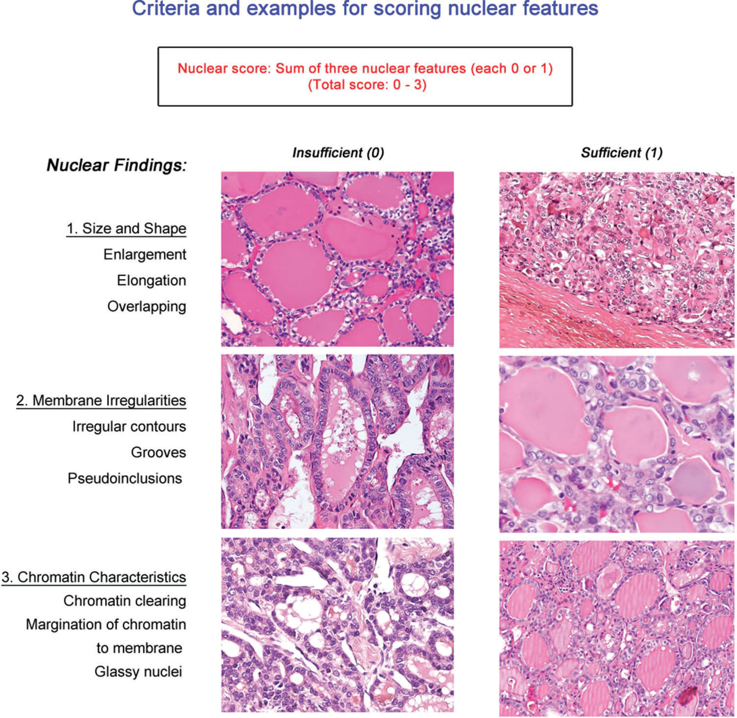
This is also true for NIFTP (Non-Invasive Follicular Thyroid Neoplasm with Papillary-like Nuclear Features). Diagnosing NIFTP requires both complete encapsulation and a detailed assessment of nuclear features, which are scored systematically on permanent sections. This level of detail is impossible to achieve on a frozen section.
4. Recognizing "Clear Benign" Patterns
Just as important as identifying malignancy is confidently recognizing benign processes, giving the surgeon the green light to proceed with a more limited resection.
- Multinodular Goiter / Colloid Nodule: These are typically easy to recognize. You will see large, variably sized follicles filled with abundant, often cracking colloid and lined by flattened, quiescent follicular cells.
- Hashimoto's Thyroiditis: The key features are a dense, diffuse lymphocytic infiltrate, often with well-formed germinal centers, and oncocytic (Hürthle cell) change in the follicular epithelium. The main pitfall is not to overcall the nuclear atypia that can be seen in Hürthle cells.
Let's look at examples of these benign entities on frozen section.
Frozen Section in Diagnosis of Thyroid Swelling
The same paper we reviewed earlier provides clear comparisons of benign conditions.
Please review Figure 2 (Colloid Goitre) and Figure 4 (Hashimoto's Thyroiditis). For each, compare the frozen section image (A) with the final H&E (B). Notice how the key architectural features (large follicles in goiter, lymphocytic infiltrate in thyroiditis) are readily apparent even on the frozen section.
By recognizing these classic benign patterns, you provide definitive, valuable information that directly impacts surgical management.
Conclusion
Interpreting a thyroid frozen section is a skill that blends pattern recognition with a deep understanding of the test's inherent limitations. Your role is not just to make a call, but to communicate the level of certainty to the surgeon.
Key Takeaways:
- Acknowledge Artifacts, Leverage Cytology: Always be aware of freezing artifacts. Use scrape or touch preps to get the best possible view of nuclear features, which are critical for diagnosis.
- Recognize Classic PTC: Confidently identify classic PTC by its papillary architecture and the constellation of nuclear features, confirming the diagnosis and enabling a one-stage total thyroidectomy.
- Defer Follicular Lesions: The most important "non-diagnosis" is recognizing an encapsulated follicular-patterned lesion and deferring the final call to permanent sections. It is the standard of care and protects the patient from both over- and under-treatment.
- Identify Clear Benign Patterns: Swiftly recognizing entities like colloid goiter or Hashimoto's thyroiditis provides equally valuable information to the surgeon.
This lesson concludes our module on gross examination and intraoperative consultation. You now have a framework for both the gross bench and the cryostat.
In our next module, we will begin our deep dive into the histopathology of permanent sections, starting with the full spectrum of Non-Neoplastic Conditions and Benign Mimics, building on the patterns you've learned to recognize today.