Introduction
Welcome back. In our last lesson, we established the critical role of the rotator cuff as a dynamic stabilizer. You learned that its health is essential not just for preventing injury, but for allowing prime movers like the deltoids and pecs to express their full strength by keeping the humeral head securely centered. We validated coaching cues like "retract the scapula" by examining the underlying biomechanics.
Today, we shift from theory to diagnostics. The learning outcome for this lesson is to analyze common shoulder training faults (e.g., impingement patterns, poor scapular mechanics) using anatomical principles. We'll learn to identify what "bad form" actually looks like from a mechanical perspective and trace it back to its anatomical roots. Think of the last lesson as understanding the design of a stable system; this lesson is about learning to recognize its failure modes.
1. Scapulohumeral Rhythm: The Foundation of Healthy Movement
Before we can identify faults, we must understand correct movement. The shoulder is a complex system where the humerus (arm bone) and the scapula (shoulder blade) must move in a synchronized dance. This coordination is called scapulohumeral rhythm.
For every 3 degrees of arm elevation (lifting your arm up), the glenohumeral (GH) joint contributes 2 degrees of movement, and the scapulothoracic (ST) joint contributes 1 degree. This 2:1 ratio is crucial. As your arm goes up, your scapula must rotate upwards to keep the glenoid socket positioned correctly under the head of the humerus, maintaining stability and creating space.

When this rhythm is disrupted, we call it scapular dyskinesis. This isn't a specific injury, but rather a term for faulty movement patterns of the scapula, which are the root cause of many common shoulder training faults.
Let's watch a short video that introduces this concept and begins to connect scapular position to potential problems.
Your Shoulder Pain Is From THIS (Not What You Think)
The video 'Your Shoulder Pain Is From THIS' by Squat University provides an excellent introduction to scapulohumeral rhythm and how its disruption can lead to pain and injury. It also introduces the key muscles that control scapular position.
Watch the first three sections of the video: Introduction (00:00 - 00:49): This sets the stage by introducing scapulohumeral rhythm. The 'Scapular X' (01:00 - 02:32): Pay close attention to the force couple between the pec minor and lower trap, which controls scapular tilt. Rhomboids and Serratus Balance (02:32 - 03:28): Focus on the relationship between these muscles, which controls scapular rotation and its distance from the spine.
This video introduces a key idea: shoulder problems often stem from an imbalance between muscles that pull the scapula in different directions. A tight pec minor and weak lower trap can cause the scapula to tilt forward, while tight rhomboids and a weak serratus anterior can prevent it from rotating upward properly. These imbalances are the direct anatomical causes of the faults we will now analyze.
2. Analyzing Common Scapular Faults (Dyskinesis)
Let's get more specific. When a lifter performs an exercise, these muscular imbalances manifest as observable movement errors. A highly detailed video from Physiotutors will help us categorize these faults systematically.
Scapular Alignment and possible causes for dyskinesia
The video 'Scapular Alignment and possible causes for dyskinesia' by Physiotutors provides a clinical breakdown of different types of scapular misalignment. It precisely identifies which muscles are typically shortened (tight) or lengthened (weak) for each fault.
Watch the section from 03:24 to 06:21. This is the core of the video. As you watch, focus on creating a mental map for each type of dyskinesia: Downward Rotated Scapula: What does it look like? Which muscles are short/tight? Which are long/weak? Winging Scapula: What is its key visual sign? Which muscle is implicated? Tilted Scapula: How does this differ from winging? Which muscles are the primary cause?
Let's synthesize this information into a table you can use for diagnosis. This connects the visual fault to its anatomical cause.
| Common Fault | What It Looks Like During Movement | Primary Anatomical Cause (Muscular Imbalance) |
|---|---|---|
| Insufficient Upward Rotation | During a lateral raise or overhead press, the top of the shoulder blade doesn't rotate up and out. The lifter may "shrug" to compensate. | Tight/Overactive: Rhomboids, Levator Scapulae, Pectoralis Minor. Weak/Inhibited: Serratus Anterior, Lower & Upper Trapezius. |
| Excessive Elevation (Shrugging) | The entire shoulder girdle lifts towards the ear, especially at the start of a lateral raise or end of a row. | Tight/Overactive: Upper Trapezius, Levator Scapulae. Weak/Inhibited: Lower Trapezius (can't depress the scapula). |
| Scapular Winging | The inside border of the shoulder blade lifts off the rib cage, especially during a push-up or bench press. | Weak/Inhibited: Serratus Anterior. |
| Anterior Tilting | The front of the shoulder rolls forward and the bottom of the scapula lifts off the back. Noticeable during the lowering phase of a row or start of a press. | Tight/Overactive: Pectoralis Minor, Short head of Biceps. Weak/Inhibited: Lower Trapezius, Serratus Anterior. |
This is the "why" behind bad form. A coach who says "stop shrugging" is observing a symptom. An evidence-based coach understands the shrug is compensation for a failure of the lower trapezius and serratus anterior to do their jobs.
3. Impingement Patterns: When Faulty Mechanics Cause Pain
Now we connect the fault to the consequence. Shoulder impingement is the painful pinching of soft tissues (usually the supraspinatus tendon and the subacromial bursa) between the head of the humerus and the acromion process of the scapula.
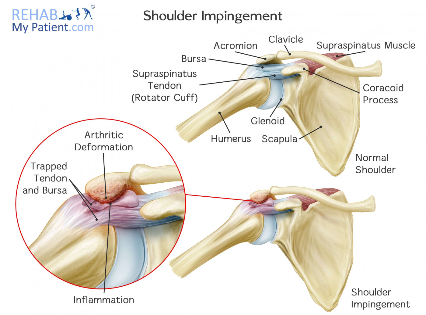
Impingement isn't random; it's a predictable outcome of poor mechanics.
Fault: Pressing in the Frontal Plane
One of the most common training faults leading to impingement is performing overhead presses with the elbows flared out to the sides (in the frontal plane). Your anatomy dictates a safer path. The glenoid socket naturally faces about 30-35 degrees forward of the frontal plane. This is called the scapular plane.
A Pro's Guide to Muscle Mechanics: The Shoulders
The article 'A Pro's Guide to Muscle Mechanics: The Shoulders' from ACE Fitness explains this concept perfectly. It will clarify why exercise selection and technique are not arbitrary.
Read the sections 'Understanding Movement of the Shoulder Joint' and 'Overhead Presses and Front Raises'. Pay special attention to the 'Try this' box, which provides a practical demonstration of the difference between the frontal and scapular planes.
As the article explains, pressing in the scapular plane provides more room for the humerus to move without jamming the rotator cuff tendons against the acromion. This is a direct anatomical principle you can apply to make exercises like the dumbbell overhead press safer and more effective.
Fault: Insufficient Scapular Upward Rotation
This is the most direct cause of impingement during overhead movements. If the scapula fails to rotate upwards as the arm elevates, the acromion effectively forms a "roof" that the humerus crashes into.
Let's examine some clear examples of dysfunctional movement in action.
How to properly & permanently resolve scapular dyskinesis
The article 'How to properly & permanently resolve scapular dyskinesis' contains excellent illustrations of faulty movement patterns. We will focus on the section that analyzes these dysfunctions.
Read two key sections in this in-depth article: Start at the heading 'Why improper scapular mechanics cause injury'. Read this short section to solidify your understanding of how downward rotation leads directly to impingement (Figure 17 is a great illustration). Next, jump to the heading 'Dysfunctional scapular movement'. Read this section carefully. It provides annotated images (Figures 26, 27, 28) showing real-world examples of scapular dyskinesis during abduction and pressing, explaining exactly what is wrong from an anatomical standpoint. Compare what you see in the images to the table we created earlier.
The article powerfully illustrates how a failure to upwardly rotate the scapula during abduction, or a tendency for the scapula to be pulled down during a press, directly leads to impingement and dysfunctional, weak movement.
Conclusion
In this lesson, you've moved from a theoretical understanding of shoulder stability to the practical skill of diagnosing instability in action. You can now analyze common training faults not as simple mistakes, but as predictable consequences of underlying anatomical and muscular imbalances.
Key Takeaways:
- Scapulohumeral Rhythm: The healthy 2:1 ratio of movement between the humerus and the scapula is the foundation of safe overhead motion.
- Scapular Dyskinesis: This is the clinical term for faulty scapular mechanics, which are the root cause of most non-traumatic shoulder injuries in the gym.
- Common Faults & Their Causes: You learned to identify key faults like insufficient upward rotation, winging, and anterior tilt, and link them to specific tight or weak muscles (e.g., weak serratus anterior, tight pec minor).
- Impingement Mechanism: You can now explain how impingement occurs—specifically, how failing to upwardly rotate the scapula or pressing in the frontal plane closes the subacromial space, pinching the rotator cuff tendons.
Preview for the Next Lesson
You have now developed the crucial skill of analysis. You can watch a movement, identify a fault, and understand its anatomical cause. The next logical step is correction. In our next lesson, you will learn to formulate anatomical coaching cues to correct common shoulder training mistakes and improve exercise execution. We will explore how to use targeted cues and exercise modifications to fix the very problems you learned to diagnose today.