Hello! Welcome to the first lesson in our course on the neuroscience of Parkinson's disease.
To understand what goes wrong in a neurological disorder, we first need to understand the fundamental building blocks of the nervous system. This lesson is dedicated to exactly that. We will explore the structure and function of the neuron, the specialized cell responsible for transmitting information throughout your brain and body.
By the end of this lesson, you will be able to describe the main parts of a neuron—the dendrites, soma, axon, and axon terminals—and explain the specific role each part plays. This knowledge is the essential foundation upon which we will build our understanding of brain circuits, neurotransmitters, and ultimately, the changes that occur in Parkinson's disease.
Let's begin by getting a general overview of what neurons are and why they are so special.
10-Minute Neuroscience: Neurons
This short video from the Neuroscientifically Challenged channel provides an excellent introduction to neurons as the information-processing units of the nervous system.
Please watch the beginning of the video, from the start until timestamp 02:12. Pay attention to how neurons use both electrical and chemical signals to communicate.
As the video mentioned, while there are over 1000 different types of neurons, most share the same fundamental components. We will now explore a "typical" neuron's structure in more detail.
The Anatomy of a Neuron: A Blueprint for Communication
Think of a neuron as a highly specialized information processing unit. It has four main parts, each with a distinct function: receiving, integrating, conducting, and transmitting information.
Here is a diagram that shows these different parts. We will refer back to this as we discuss each component.
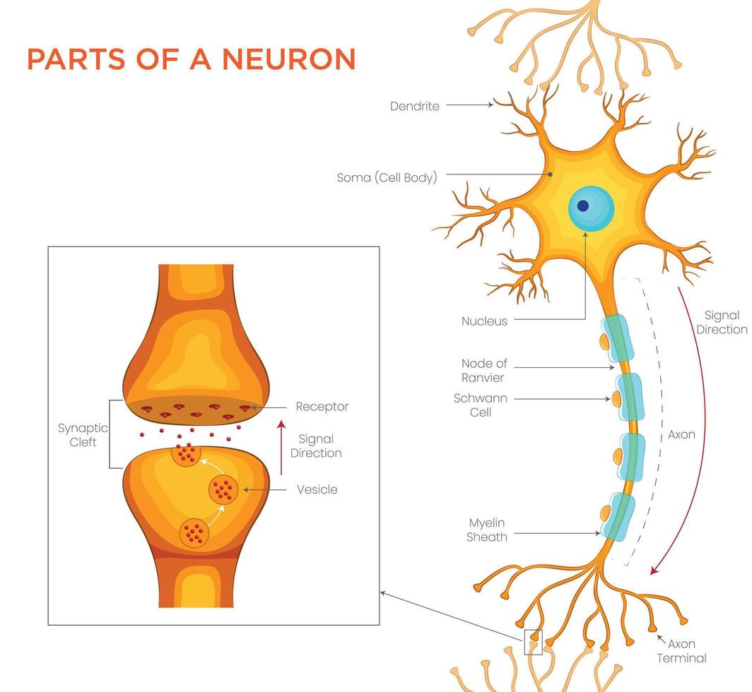
Now, let's break down each of these parts.
1. Dendrites: The Input Receivers
The dendrites are the tree-like extensions that branch out from the main cell body. Their primary function is to receive signals from other neurons.
- The surface of dendrites is covered with thousands of specialized proteins called receptors.
- When chemical messengers called neurotransmitters are released by a neighboring neuron, they bind to these receptors.
- This binding event triggers a small electrical change in the dendrite.
You can think of the dendrites as the neuron's "inbox" or "listening antennae," constantly collecting messages from the surrounding neural network.
Parts of a neuron: Structure and functions
For a more detailed textual description, let's look at the Kenhub article 'Parts of a neuron'.
Please read the section titled 'Dendrites'. Note the mention of 'dendritic spines' and how they increase the surface area available for receiving signals.
2. The Soma: The Integration and Metabolic Center
The signals received by the dendrites all travel to the soma, or cell body. The soma has two critical jobs:
- Metabolic Center: It's the neuron's life-support system. It contains the nucleus (which holds the cell's DNA) and other organelles responsible for producing energy and manufacturing the proteins necessary for the neuron's survival and function.
- Integration Center: The soma sums up all the incoming signals—both excitatory (go!) and inhibitory (stop!)—from the dendrites. This process happens at a specialized region where the soma joins the axon, called the axon hillock. If the total signal crosses a certain threshold, the neuron "decides" to fire its own signal. This is a fundamental aspect of neural computation.
Parts of a neuron: Structure and functions
The Kenhub article provides a clear summary of the soma's dual roles.
Read the section titled 'Cell body'. Pay close attention to the descriptions of the organelles and the function of the axon hillock.
3. The Axon: The Conduction Cable
If the integrated signal at the axon hillock is strong enough, the neuron fires an electrical impulse called an action potential. This signal is sent down the axon, a long, slender projection that acts like a biological electrical wire.
- The axon's job is to conduct the action potential, often over long distances, without the signal degrading.
- Many axons are wrapped in a fatty insulating layer called the myelin sheath. This insulation dramatically speeds up signal transmission.
A crucial function that isn't always highlighted is axonal transport. Since the axon can be very long and lacks the machinery to make its own proteins, the soma must ship essential materials (like neurotransmitters, proteins, and organelles like mitochondria) down the axon. This process also works in reverse to transport waste materials and signaling molecules back to the soma.
Neurology | Neuron Anatomy & Function
The following video from Ninja Nerd offers a fantastic and detailed explanation of the axon, including a deep dive into the fascinating process of axonal transport.
Please watch the section from 12:56 to 22:57. Focus on understanding the difference between anterograde (forward) and retrograde (backward) transport and the clinical examples given (like how viruses can hijack this system).
4. Axon Terminals: The Output Transmitters
The axon ends by branching into several axon terminals (also called terminal boutons). This is the output end of the neuron. When the action potential arrives at the axon terminal, it triggers the release of neurotransmitters into a tiny gap called the synapse.
This process allows the neuron to transmit its signal to the next cell in the circuit, which could be another neuron, a muscle cell, or a gland.
Neurology | Neuron Anatomy & Function
Let's return to the Ninja Nerd video to see exactly how this transmission happens at the axon terminal.
Watch from 22:57 to 28:22. This section clearly explains how the arrival of an action potential leads to neurotransmitter release. The example of SSRIs is a great illustration of how understanding this basic process is key to pharmacology.
Test your understanding!
Imagine a neuron is a single-person company that processes information and sends out memos. Match the parts of the neuron to the following company roles:
- Neuron Parts: Dendrites, Soma, Axon, Axon Terminal
- Company Roles:
- The CEO's office, where all incoming mail is read, decisions are made, and the company is maintained.
- The mailroom, which receives all incoming letters and packages.
- The courier service, responsible for delivering memos over long distances to other offices.
- The shipping department, which packages and sends the memos out.
Show answer
- Dendrites -> 2. The mailroom (receives inputs).
- Soma -> 1. The CEO's office (integrates information, makes decisions, and maintains the cell).
- Axon -> 3. The courier service (conducts the signal).
- Axon Terminal -> 4. The shipping department (transmits the signal to the next cell).
Conclusion
In this lesson, we have dissected the neuron and seen how its structure is perfectly suited for its function as the nervous system's primary communicator.
Here are the key takeaways:
- Dendrites act as the receptive zone, collecting signals from other neurons.
- The Soma (or cell body) serves as both the metabolic center and the integration hub, where it sums up inputs to decide whether to fire.
- The Axon is the conductive cable that carries the electrical signal (action potential) away from the cell body.
- Axon terminals form the transmissive zone, releasing chemical neurotransmitters to communicate with the next cell.
Understanding this basic unit is the first and most critical step in our journey. All the complex functions of the brain, from thought to movement, arise from the coordinated activity of billions of these cells. In Parkinson's disease, the progressive loss of a specific type of neuron—dopaminergic neurons—disrupts this communication, leading to the symptoms we will explore later.
In our next lesson, we will zoom in on the axon and explore the fascinating electrical event at the heart of neural communication: the action potential. We will see how ions moving across the neuron's membrane create the very signal that travels from the soma to the axon terminal.