Hello! In our last lesson, we established the crucial concept of osmolality—the measure of solute concentration that creates the potential for water to move between compartments. We now know that the body maintains a precise plasma osmolality of 280-295 mOsm/kg, primarily dictated by sodium.
Today, we will explore how this potential is translated into actual fluid movement. Your learning outcome for this lesson is to explain how osmotic and hydrostatic pressures govern water movement between fluid compartments. We will unpack the two fundamental forces—the "push" of hydrostatic pressure and the "pull" of osmotic pressure—that dictate the distribution of water throughout your body. Understanding this dynamic interplay is the foundation for comprehending everything from normal tissue hydration to the pathophysiology of conditions like edema and the principles of intravenous fluid therapy.
The Two Major Forces: Push and Pull
Water moves between compartments for two main reasons: either it is being pushed or it is being pulled. In physiology, these pushes and pulls are given specific names: hydrostatic pressure and osmotic pressure.
1. Hydrostatic Pressure: The "Push"
This is the pressure exerted by a fluid at rest due to the force of gravity or, more importantly in our case, the pressure exerted by blood within the circulatory system. Think of it as the mechanical force of the fluid against the walls of its container. In the body, the primary source of hydrostatic pressure in the vascular system is the pumping action of the heart. This pressure is what tends to push fluid out of the blood vessels and into the surrounding tissues.
2. Osmotic Pressure: The "Pull"
This is the force that "pulls" water across a semipermeable membrane. It's generated by the presence of solutes that cannot easily cross the membrane. As we discussed in the last lesson, water will always move from an area of lower solute concentration (higher water concentration) to an area of higher solute concentration (lower water concentration) in an attempt to dilute the solutes and achieve equilibrium. This "pull" exerted by the solutes on water is the osmotic pressure.
A particularly important type of osmotic pressure in the blood vessels is oncotic pressure (or colloid osmotic pressure). This is the osmotic pressure specifically generated by large molecules, primarily the plasma protein albumin. Because albumin is too large to easily pass through the capillary walls, it stays within the blood vessels and exerts a constant osmotic pull, drawing fluid into the capillaries from the interstitial space.
The following reading provides formal definitions of these concepts.
Physiology, Body Fluids - StatPearls - NCBI Bookshelf
Please read the section "Mechanism" in this StatPearls article.
Focus on the paragraph that begins Fluid moves throughout. It defines both the osmotic "pull" and the hydrostatic "push," setting the stage for understanding their combined effect. Don't worry about the Starling equation just yet; we will break that down next.
Now that we have defined our two primary forces, let's see how they work together in the body's different fluid compartments.
Fluid Exchange in the Capillaries: The Starling Forces
The movement of fluid between the plasma in the capillaries and the surrounding interstitial fluid is a dynamic process governed by the balance between hydrostatic and oncotic pressures on both sides of the capillary wall. These are collectively known as Starling's forces.
There are four key forces to consider:
- Capillary Hydrostatic Pressure (Pc): Pushes fluid out of the capillary.
- Capillary Oncotic Pressure (πc): Pulls fluid into the capillary.
- Interstitial Hydrostatic Pressure (Pi): Pushes fluid into the capillary.
- Interstitial Oncotic Pressure (πi): Pulls fluid out of the capillary.
The video below by The Noted Anatomist provides an excellent visual explanation of how these forces interact.
Starling Forces: Hydrostatic vs Oncotic Pressures and Edema
This video, "Starling Forces," breaks down each component of capillary fluid exchange. First, watch the introduction and the section on hydrostatic pressure, from the beginning to the end of the edema explanation. Notice how hydrostatic pressure is highest at the arterial end and decreases towards the venous end. Next, watch the segment on oncotic pressure, from the start of the section until the "net filtration pressure" section begins. Pay attention to why albumin creates a constant "sucking force." Finally, watch the segment on net filtration pressure, from the start of this section to the discussion of the lymphatic system. Here, the presenter puts it all together with a numerical example using Starling's equation.
The video provides a clear, step-by-step breakdown of the forces. Focus on grasping the conceptual "tug-of-war" between the pushing hydrostatic pressure and the pulling oncotic pressure.
As the video explains, the balance of these forces changes along the length of the capillary.
- At the arterial end: Capillary hydrostatic pressure is high (around 35 mm Hg), overpowering the constant capillary oncotic pressure (around 25 mm Hg). The net result is filtration, where fluid and small solutes are pushed out of the capillary into the interstitial space.
- At the venous end: As blood flows through the capillary, pressure drops. Here, the capillary hydrostatic pressure is lower (around 18 mm Hg), and it is now weaker than the capillary oncotic pressure. The net result is reabsorption, where fluid is pulled back into the capillary from the interstitial space.
Overall, filtration slightly exceeds reabsorption. About 20 liters of fluid are filtered out of capillaries each day, while about 17-18 liters are reabsorbed. What happens to the extra 2-3 liters of fluid left in the interstitial space? It is collected by the lymphatic system and eventually returned to the bloodstream, preventing the continuous accumulation of fluid in the tissues.
Fluid Exchange Between ECF and ICF: It's All About Osmosis
When we consider the movement of fluid between the interstitial fluid (part of the ECF) and the intracellular fluid (ICF), the rules change slightly. While capillary walls are leaky, cell membranes are highly selective. Crucially, hydrostatic pressure differences across the cell membrane are negligible. Therefore, water movement between the ECF and ICF is driven almost exclusively by osmotic gradients.
Because cell membranes contain water channels (aquaporins), water moves freely to equalize the osmolality between the inside and outside of the cell. Sodium (Na⁺), the main ECF cation, is actively pumped out of cells by the Na⁺-K⁺-ATPase and is effectively restricted to the ECF. This makes sodium the primary determinant of ECF osmolality and the main driver of water movement across the cell membrane.
- If the ECF becomes hypertonic (higher osmolality, e.g., from dehydration or high salt intake), water will be pulled out of the cells into the ECF, causing the cells to shrink (crenate).
- If the ECF becomes hypotonic (lower osmolality, e.g., from drinking excessive amounts of water), water will be pulled from the ECF into the cells, causing them to swell. This can be very dangerous, especially in the brain.
The following video provides a clear, practical example of how this works.
Fluid Compartments & Fluid Balance
Watch this clip from "Fluid Compartments & Fluid Balance" by Dr Matt & Dr Mike.
Focus on the segment from when the presenter discusses insensible water loss to the end of the cellular dehydration example. This demonstrates perfectly how losing more water than salt from the ECF concentrates the interstitial sodium, pulling water out of the cells.
Clinical Application: The Pathophysiology of Edema
You are now equipped to understand the mechanisms behind edema, the clinical term for swelling caused by excess fluid trapped in the body's tissues. Edema is a direct consequence of disrupted Starling forces.
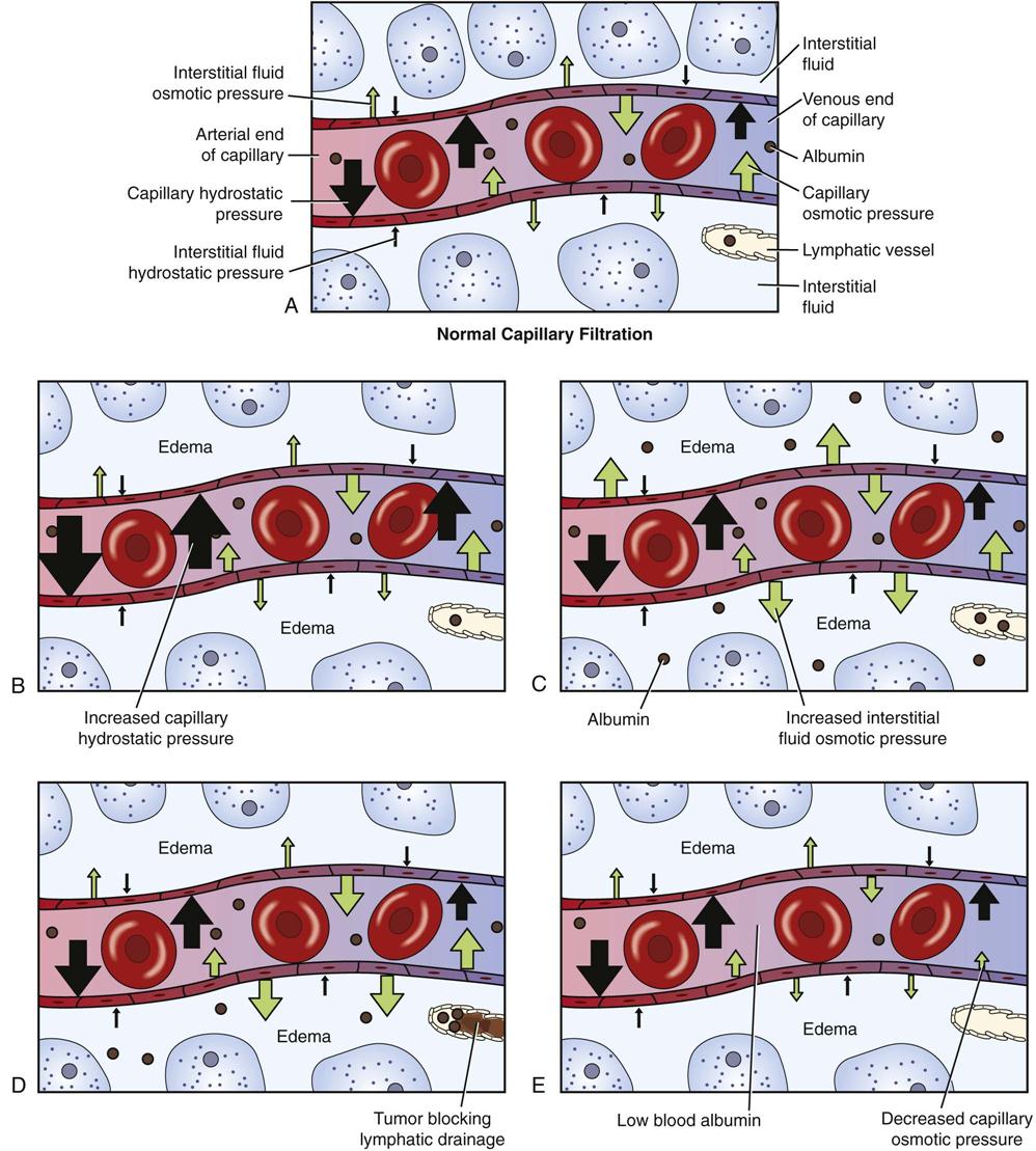
Let's break down the common causes based on the principles we've learned:
- Increased Capillary Hydrostatic Pressure (Pc): If pressure in the capillaries increases, more fluid is pushed out. A classic example is congestive heart failure. When the right side of the heart fails to pump effectively, blood backs up in the systemic venous circulation, increasing venous and capillary pressure, leading to peripheral edema (e.g., swollen ankles).
- Decreased Capillary Oncotic Pressure (πc): If the concentration of plasma proteins, especially albumin, falls, the "pulling" force that holds fluid inside the capillaries is reduced. This is common in liver disease (since the liver synthesizes albumin), nephrotic syndrome (where large amounts of albumin are lost in the urine), and severe malnutrition. The result is a net movement of fluid into the interstitial space.
- Increased Capillary Permeability: In conditions like inflammation, burns, or sepsis, the capillary walls become "leaky." Plasma proteins can then escape into the interstitial fluid, increasing the interstitial oncotic pressure (πi). This both reduces the capillary oncotic pressure and increases the osmotic pull into the interstitium, causing significant edema.
- Lymphatic Obstruction: If the lymphatic system is blocked or damaged (e.g., from surgery, infection, or a tumor), it cannot drain the excess interstitial fluid. This leads to a type of edema called lymphedema.
Physiology, Body Fluids - StatPearls - NCBI Bookshelf
Now, please read the section "Clinical Significance" in the same StatPearls article.
Read the paragraphs explaining pulmonary edema and edema in liver disease. Note how the authors directly attribute these clinical conditions back to imbalances in the Starling equation.
Conclusion
In this lesson, we have dissected the fundamental forces that govern all fluid movement in the body. You should now be able to explain the "why" behind fluid shifts, from the microscopic level of a single capillary to the systemic presentation of edema.
Here are the key takeaways:
- Fluid movement is driven by two main forces: hydrostatic pressure (a "pushing" force) and osmotic pressure (a "pulling" force).
- Across capillaries, the balance of four Starling forces (hydrostatic and oncotic pressures in both the capillary and interstitium) determines whether filtration or reabsorption occurs.
- Across cell membranes, water movement is dictated almost entirely by osmotic gradients, as water moves to equalize the osmolality between the ICF and ECF.
- Clinical conditions like edema arise from specific disruptions in the balance of Starling forces, such as increased hydrostatic pressure or decreased oncotic pressure.
You have now completed the foundational module on body fluids. You understand where the water is, how its concentration is measured, and the physical principles that govern its movement. In our next module, we will build on this foundation to explore how the body actively regulates water balance through hormonal and neural systems, starting with the thirst mechanism and the critical role of Antidiuretic Hormone (ADH).