Welcome back. In the previous lesson, you distinguished a plausible Ebola exposure from mere proximity by looking for a source of virus, infectious material, and a way into the body. Now that exposure history becomes one part of a different judgment: when should an illness be treated as a suspected case, and what can a diagnostic test establish?
This lesson follows a fictional patient from exposure to symptoms and testing. The central distinction is between reason to suspect Ebola disease and laboratory confirmation. A person may need prompt precautions and assessment well before a test result is available.
Suspicion begins with a history, not a distinctive symptom
Early Ebola disease can look like many other illnesses. Fever, fatigue, headache, and muscle pain may appear first; vomiting or diarrhoea can develop later. Bleeding is not required to suspect Ebola. These symptoms matter most when placed alongside when a possible exposure occurred and what the person encountered.
Watch this short segment for the diagnostic problem it presents: symptoms alone do not tell a clinician whether the cause is Ebola, malaria, or another infection.
Ebola - Transmission, Symptoms, Diagnosis, and Treatment
Strong Medicine’s “Ebola - Transmission, Symptoms, Diagnosis, and Treatment” connects the early clinical picture to exposure history and introduces the confirmatory test.
Watch symptoms and history for why ordinary-looking symptoms can still warrant suspicion after a relevant exposure. Then watch RT-PCR diagnosis for what the blood test detects. Keep the time since symptom onset in mind; we will use it to interpret a negative result.
A useful initial account has two timelines. The exposure timeline establishes whether a plausible encounter with infectious material occurred within the possible incubation period of 2–21 days before illness. The symptom timeline records when the first compatible symptom began. They answer different questions: exposure timing helps establish suspicion, while symptom timing helps interpret testing.
This illustrative flowchart shows why a symptomatic person with a relevant history is assessed promptly rather than told to wait for a laboratory result.
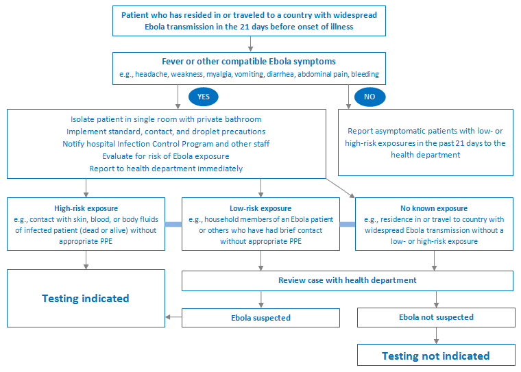
The chart starts with travel or residence in an area of widespread transmission. In an actual outbreak, investigators also consider other relevant histories, such as contact with a symptomatic patient or infectious animal material. Even within the chart, no known direct exposure is not an automatic reason to dismiss compatible illness: the case is reviewed with the health department. Which histories meet a formal suspected-case definition depends on the outbreak setting and current public-health guidance.
What the blood test detects—and why timing matters
The main molecular method used to confirm acute Ebola disease is RT-PCR, a form of nucleic acid amplification testing. It looks for viral RNA in a patient sample, usually blood. A properly validated positive result in a suspected, symptomatic patient can confirm infection; the assay used determines whether the result identifies a particular virus species or requires further testing to do so. RT-PCR is not a test for “how sick someone looks,” and a test result must be interpreted alongside the clinical and exposure history.
First, read the World Health Organization’s guidance on who should be tested and why a negative test taken very early can be misleading.
Diagnostic testing for Ebola disease and Marburg virus disease
The World Health Organization’s diagnostic guidance gives the testing rule that will anchor the case below. It also explains why clinicians investigate other possible causes of the same symptoms.
In Section 3, “Indications for testing and importance of differential diagnosis and coinfections” (pp. 5–6), read the timing guidance, beginning with the paragraph about an initial negative blood sample and ending with the paragraph about monitoring contacts. Then read the differential-diagnosis explanation, from the paragraph beginning “Given the non-specific clinical presentation” through its examples of malaria and bacterial infection. Focus on the difference between an asymptomatic exposed person and a symptomatic suspected case.
Viral RNA in blood generally becomes detectable around symptom onset, but detection may not yet be reliable in the earliest phase. The WHO guidance therefore makes a practical distinction:
- Before symptoms: a negative test cannot rule out an infection that may become detectable later. Routine laboratory testing of asymptomatic contacts is not recommended; they are monitored for symptoms after their last exposure.
- After symptoms begin: testing is appropriate when someone meets the suspected-case criteria. If the first blood sample is negative and was collected within 72 hours of symptom onset, a new sample should be tested more than 72 hours after onset.
The second sample is not a repeat for its own sake. It addresses a specific limitation: the first sample may have been collected before enough viral RNA was detectable in blood.
Diagnostic testing for Ebola disease and Marburg virus disease
Continue with the WHO guidance for a closer look at what RT-PCR is used for during an outbreak.
In Section 5.2, “NAAT for orthoebolaviruses and orthomarburgviruses” (pp. 11–12), read the opening explanation, from the subsection’s first paragraph through the paragraph describing why NAAT is preferred. Read the immediately following sentence, beginning “It is also the only documented method,” on early detection. Treat “as early as” as a possibility, not a promise that every infection will be detectable at that hour.
A suspected case across two timelines
Consider this fictional account. On day 0, a person cleans vomit from someone who is visibly ill during an Ebola outbreak. Fluid splashes near the cleaner’s eye. On day 9, the cleaner develops fever, fatigue, and headache, without bleeding. A blood sample taken on day 10 is negative by RT-PCR.
The day-0 encounter is a plausible exposure, and symptom onset nine days later falls within the 2–21-day incubation period. Fever and headache do not establish Ebola; they also do not become reassuring because there is no bleeding. The history makes this a case for prompt professional assessment, appropriate infection precautions, and testing under the local outbreak protocol.
Now locate the negative result on the symptom timeline. The day-10 sample was taken roughly one day after symptoms began, within the first 72 hours. It does not reliably settle the question. Under the WHO guidance, a new blood sample should be collected more than 72 hours after onset if this remains a suspected case. Suppose that sample is collected on day 13 and a validated species-specific RT-PCR is positive. That later result confirms infection with the species the assay identifies; it also explains why the earlier negative result could not safely close the investigation.
Testing does not replace care while the answer is uncertain. Clinicians can assess and treat other plausible illnesses, including malaria where relevant, while maintaining the precautions warranted for a suspected Ebola case. Finding another infection does not, by itself, establish that Ebola is absent: coinfections can occur.
Reading results without giving them more certainty than they have
Timing is a major reason a negative result may mislead, but not the only one. Sample quality, handling, transport, and test performance also affect whether viral RNA is detected. The WHO guidance describes these limitations alongside its interpretation of positive results.
Diagnostic testing for Ebola disease and Marburg virus disease
Finish with the WHO’s interpretation guidance to see why a laboratory result is considered together with the patient’s history.
In Section 6, “Interpretation of laboratory results” (p. 15), read the confirmation paragraph, from its opening sentence through the description of positive species-specific testing. Then read the false-negative factors in the next paragraph. Focus on why a result is evidence to interpret, rather than a substitute for knowing when and how the sample was collected.
For an asymptomatic contact, a negative RT-PCR result is not a certificate that infection will not develop; symptom monitoring remains the relevant response. For a symptomatic suspected case tested very early, a negative result calls for follow-up testing at the recommended time, not immediate dismissal. A negative result from a properly collected sample after the early testing window weighs much more strongly against acute Ebola disease, though clinicians and public-health teams still interpret it in context. A positive result requires attention to which virus the assay actually detects—an issue that matters when outbreak reports name a particular Ebola virus.
These distinctions also explain why the first response to a concerning presentation is not “wait for proof.” Precautions and professional assessment address the risk during the period when the diagnosis is still uncertain.
The judgment to carry forward
A suspected Ebola case is interpreted by combining a credible exposure history, compatible but nonspecific symptoms, and two dates: last possible exposure and symptom onset. RT-PCR looks for viral RNA in blood, but a negative result before symptoms—or very soon after they begin—cannot answer the same question as an appropriately timed test. If an initial sample is negative within 72 hours of symptom onset, the WHO recommends a new sample more than 72 hours after onset.
In the next lesson, the focus moves from recognizing illness to understanding how supportive care addresses fluid loss and its effects on the body.