Hello! Welcome to your next lesson on Coagulation Studies.
In our last session, we focused on how to interpret D-dimer results, primarily for ruling out venous thromboembolism (VTE) in low-risk patients. We briefly mentioned that D-dimer is also a crucial marker in a much more dramatic condition: Disseminated Intravascular Coagulation (DIC).
Today, we will dive deep into that topic. Your learning outcome is to recognize the classic coagulation pattern of Disseminated Intravascular Coagulation (DIC). DIC is a life-threatening syndrome that you will encounter in critically ill patients. It presents a paradox: a disease of widespread clotting that ultimately leads to catastrophic bleeding. Recognizing the laboratory "fingerprint" of DIC is a vital clinical skill.
1. What is DIC? A Thrombo-Hemorrhagic Catastrophe
Disseminated Intravascular Coagulation is not a disease in itself, but rather an acquired syndrome triggered by a severe underlying illness. Think of it as the coagulation system going haywire on a systemic level.
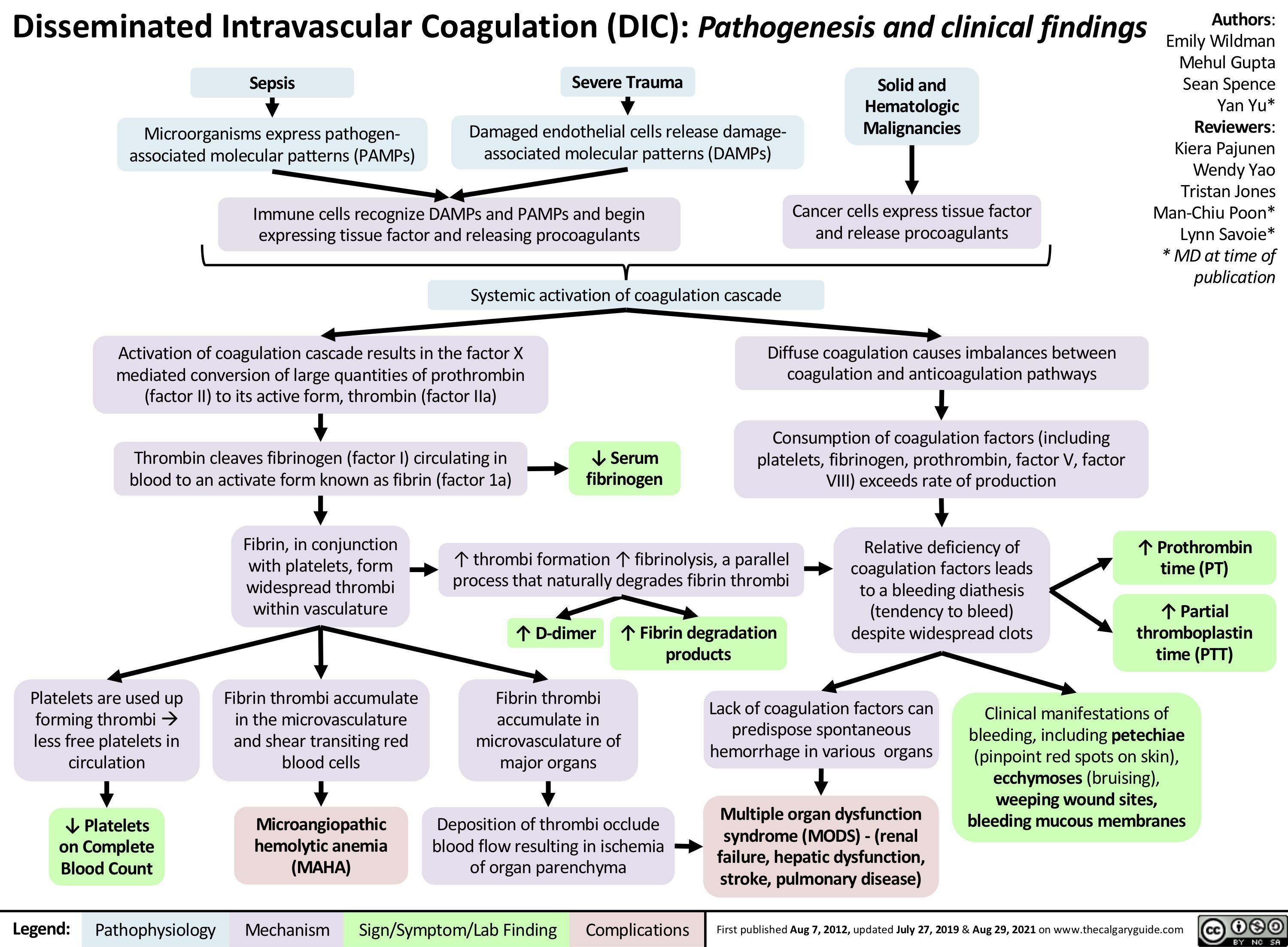
A massive trigger—like widespread infection or major trauma—causes a storm of pro-inflammatory cytokines and the release of tissue factor into the circulation. This sets off a massive, uncontrolled activation of the coagulation cascade throughout the body's small blood vessels.
This process has two devastating consequences that occur almost simultaneously:
- Thrombosis: Thousands of microthrombi (tiny clots) form in the microvasculature, obstructing blood flow and causing ischemic end-organ damage (e.g., kidney, lung, and brain injury).
- Consumption & Bleeding: The body uses up its platelets and clotting factors faster than they can be produced in the process of making these widespread clots. This "consumptive coagulopathy" leaves the patient with no ability to form clots where they are actually needed, leading to severe, uncontrollable bleeding.
The flowchart below provides a great overview of this entire process, from triggers to clinical consequences.
2. Clinical Triggers: When to Suspect DIC
Because DIC is a secondary process, it's crucial to recognize the clinical settings in which it commonly occurs. If you see a patient with one of these conditions who starts to bleed unexpectedly (e.g., oozing from IV sites, hematuria), your suspicion for DIC should be very high.
Common triggers for DIC include:
- Sepsis (especially from gram-negative bacteria)
- Trauma (especially severe crush injuries or neurotrauma)
- Obstetric complications (e.g., amniotic fluid embolism, placental abruption)
- Pancreatitis (severe acute pancreatitis)
- Malignancy (especially adenocarcinomas and acute promyelocytic leukemia - APL)
- Nephrotic syndrome, severe viral infections, Transfusion reactions, and snakebites are other causes.
A common mnemonic is STOP Making New Thrombi.
Coagulopathy | Clinical Medicine
To understand the 'why' behind DIC, watch this segment from Ninja Nerd. He clearly explains how triggers like sepsis lead to a cytokine storm that activates the coagulation cascade systemically, resulting in the consumption of clotting factors and platelets.
Watch the video from 40:10 to 44:32. Focus on the connection between the underlying inflammatory process and the subsequent widespread activation of procoagulants and platelets.
3. The Classic Laboratory Pattern of DIC
Recognizing DIC hinges on identifying a characteristic pattern of abnormalities across several key coagulation tests. No single test is diagnostic; it's the combination that tells the story.
Here is the "classic" pattern you must learn to recognize:
| Lab Test | Expected Finding in DIC | Rationale |
|---|---|---|
| Platelet Count | Low (Thrombocytopenia) | Platelets are consumed in the formation of thousands of microthrombi. |
| PT/INR & aPTT | High (Prolonged) | Clotting factors from both extrinsic and intrinsic pathways are consumed, impairing clot formation. |
| Fibrinogen | Low | Fibrinogen (Factor I) is consumed as it's converted to fibrin to create the microthrombi. |
| D-dimer | Very High | The body's fibrinolytic system works overtime to break down the massive burden of cross-linked fibrin. |
| Peripheral Smear | Schistocytes | Red blood cells are sheared and fragmented as they try to squeeze past fibrin strands in small vessels. |
Coagulopathy | Clinical Medicine
This next clip from the same Ninja Nerd video provides a perfect walkthrough of these exact lab findings and explains the 'why' behind each one. This is the core of today's lesson.
Watch from 44:32 to 47:35. Pay close attention as he systematically breaks down each component of the DIC lab panel.
An important clinical nuance: Fibrinogen is an acute-phase reactant, meaning its levels can increase in response to inflammation. In the early stages of DIC, a patient's fibrinogen level might be "normal" or even high, but this could represent a significant drop from a much higher baseline. A falling trend in fibrinogen is often more informative than a single low value.
4. Making the Diagnosis: The ISTH Scoring System
In a real clinical scenario with a critically ill patient, the lab results can be complex. To standardize the diagnosis of overt DIC, the International Society on Thrombosis and Haemostasis (ISTH) developed a practical scoring system. This tool combines the key lab values we've just discussed.
The diagnosis requires two things:
- The patient must have an underlying disorder known to be associated with DIC.
- The lab results must meet a certain score threshold.
Disseminated Intravascular Coagulation - DIC
Please study this resource from ARUP Laboratories. It presents the ISTH Overt DIC Scoring System in a clear, easy-to-read table. This is the practical tool you would use on the wards.
Read the section 'Criteria for Diagnosis' and focus on the table titled 'ISTH Overt DIC Scoring System'. Familiarize yourself with how points are assigned for Platelet count, D-dimer, Fibrinogen, and PT.
As you can see from the scoring system, a score of ≥ 5 is compatible with overt DIC. This system formalizes the pattern recognition we are learning today.
Test your understanding!
A 65-year-old man with severe sepsis from pneumonia is in the ICU. He develops oozing from his IV lines. You suspect DIC and get the following labs:
- Platelet count: 45,000/µL
- PT: prolonged by 7 seconds
- Fibrinogen: 0.8 g/L
- D-dimer: Strong increase
Using the ISTH Overt DIC Scoring System, calculate his score. Is this compatible with overt DIC?
Show answer
Let's calculate the score:
- Platelet count < 50,000/µL = 2 points
- PT prolonged > 6 seconds = 2 points
- Fibrinogen < 1.0 g/L = 1 point
- D-dimer strongly elevated = 3 points
Total Score = 2 + 2 + 1 + 3 = 8 points.
Since the score is ≥ 5, this is highly compatible with a diagnosis of overt DIC.
5. Distinguishing DIC from Severe Liver Disease
One of the most common diagnostic dilemmas is distinguishing DIC from the coagulopathy of severe liver disease. Both conditions can present with prolonged PT/aPTT, thrombocytopenia, and bleeding. However, a few key labs can help you tell them apart.
Coagulopathy | Clinical Medicine
This final video segment explains the key differences between the lab patterns in DIC and cirrhosis. Understanding these distinctions is a higher-level skill that will be very useful in your clinical rotations.
Watch from 47:35 to 52:03. Focus on the differences in D-dimer, the presence of schistocytes, and the level of Factor VIII.
Here's a summary of the key differentiators:
- D-dimer: Markedly elevated in DIC due to massive fibrinolysis. It is typically normal or only mildly elevated in liver disease (unless the patient has a separate reason for clotting).
- Factor VIII: The liver produces most clotting factors, but Factor VIII is produced by endothelial cells. In liver failure, Factor VIII levels are normal or high. In DIC, all factors are consumed, so Factor VIII levels are low. While not always measured, this is the classic textbook distinction.
Conclusion
You have now learned to recognize the characteristic laboratory pattern of Disseminated Intravascular Coagulation. This is a critical skill for managing severely ill patients where early recognition can have a significant impact on outcomes, primarily by prompting an aggressive search for and treatment of the underlying trigger.
Key Takeaways:
- Concept: DIC is a thrombo-hemorrhagic syndrome where widespread microvascular clotting leads to a consumptive coagulopathy and subsequent bleeding.
- Triggers: Always suspect DIC in the context of sepsis, severe trauma, obstetric catastrophes, and certain malignancies.
- The Classic Pattern: The hallmark lab findings are low platelets, low fibrinogen, prolonged PT/aPTT, and a markedly elevated D-dimer. Schistocytes on the peripheral smear are also a key finding.
- Practical Diagnosis: The ISTH scoring system is a standardized tool used in clinical practice to diagnose overt DIC based on this lab pattern.
- Treatment Focus: The cornerstone of managing DIC is to aggressively treat the underlying cause.
In our next lesson, we will broaden our focus to assess bleeding risk from coagulation study results prior to invasive procedures. Your understanding of the profound coagulopathy seen in DIC serves as a powerful example of why this assessment is so critical, especially in the ICU setting.