Hello! Welcome to the first lesson in our module on coagulation studies.
In our last lesson on liver function, we saw how the INR is a critical marker of the liver's synthetic capability. This provides a perfect bridge to today's topic, as the INR is one of the two foundational tests we use to assess the entire coagulation system.
Your goal for this lesson is to learn how to interpret Prothrombin Time (PT/INR) and Partial Thromboplastin Time (PTT) to localize defects in the coagulation cascade. This skill is like having a map and a set of tools that allow you to pinpoint where a patient's clotting problem might be, guiding your differential diagnosis for bleeding or thrombosis.
The Coagulation Cascade: A Functional Map
Before we can interpret the tests, we need to understand the "map" they are based on: the coagulation cascade. This is a simplified model of the chain reaction among various clotting factors (proteins) that ultimately leads to the formation of a stable fibrin clot.
The cascade is traditionally divided into three parts:
- The Extrinsic Pathway: Activated by "extrinsic" factors outside the blood, like tissue factor, which is exposed after an injury. This is considered the primary initiator of coagulation in vivo.
- The Intrinsic Pathway: Activated by factors "intrinsic" to the blood itself, such as when blood contacts a foreign surface or specific charged particles. This pathway amplifies the clotting process.
- The Common Pathway: Where the extrinsic and intrinsic pathways converge to complete the clot formation process.
A memorable way to organize these factors and pathways is presented in the following short video.
How To FINALLY Understand The Coagulation Cascade
This video from Conan Liu, M.D. offers a quick and memorable mnemonic for learning the factors in each pathway. This will be our foundational map for the rest of the lesson.
Watch the first 1 minute and 39 seconds of the video. Focus on remembering the placement of the factors: 'X marks the spot', 'lucky number seven', and the 'dollar bills' in the common pathway.
Here is a static diagram that visualizes the same concept. It will be a useful reference as we proceed.
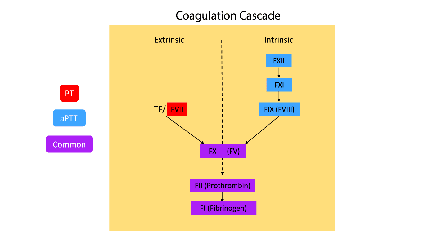
With this map in mind, let's look at the tools we use to test its integrity.
The Diagnostic Tools: PT/INR and PTT
Prothrombin Time (PT) and Partial Thromboplastin Time (PTT) are lab tests that measure how long it takes for a clot to form in a plasma sample after specific triggers are added. Each test triggers a different part of the cascade.
To understand what these tests are actually measuring in the lab, let's watch a clear explanation.
Hemostasis: Lesson 4 - Tests (INR, PTT, platelets, fibrinogen, D-dimer)
The 'Hemostasis: Lesson 4' video by Strong Medicine provides an excellent, clinically-focused explanation of how PT/INR and PTT are performed and what they measure.
Please watch from 6:34 to 13:08. As you watch, focus on answering these two questions: What is added to the patient's plasma to initiate the PT, and why does this specifically test the extrinsic pathway? What is different about the reagent added for the PTT, and why does this test the intrinsic pathway?
To summarize the key points from the video:
-
Prothrombin Time (PT/INR):
- What it measures: The integrity of the Extrinsic and Common Pathways.
- How it works: Thromboplastin (which contains tissue factor) is added to the plasma. Since tissue factor is the trigger for the extrinsic pathway, this test measures the function of Factor VII and all the common pathway factors (X, V, II, and I).
- INR: The International Normalized Ratio (INR) is just a standardized version of the PT. It allows results from different labs to be compared, which is essential for monitoring medications like warfarin. A normal INR is approximately 1.0.
-
Activated Partial Thromboplastin Time (aPTT or PTT):
- What it measures: The integrity of the Intrinsic and Common Pathways.
- How it works: An activating substance (like silica) is added, but no tissue factor. This triggers the intrinsic pathway, measuring the function of factors XII, XI, IX, and VIII, as well as the common pathway factors.
Test your understanding!
A patient has hemophilia A, a genetic disorder causing a deficiency of Factor VIII. Based on the cascade diagram, which screening test (PT/INR or PTT) do you expect to be abnormally prolonged?
Show answer
You would expect the PTT to be prolonged. Factor VIII is part of the intrinsic pathway, which is assessed by the PTT. The PT/INR, which assesses the extrinsic and common pathways, would be normal.
Localizing the Defect: Interpreting the Patterns
Now we can combine our map and our tools. By looking at which test is abnormal, we can localize the problem to a specific part of the cascade. There are three main patterns to recognize.
For a detailed breakdown of the causes for each pattern, please review the following resource. It presents a clear, organized list that you will find very useful in clinical practice.
Interpreting Coagulation Studies
The article 'Interpreting Coagulation Studies' from MedSchool.co provides a concise, high-yield summary of the causes for abnormal coagulation profiles. We will use it to build our differential for each pattern.
Read the section 'Aetiology of Coagulopathy' and pay close attention to the lists under the heading 'Causes of Abnormal Coagulation Profile'. We will walk through these patterns one by one.
Let's break down the patterns based on that reading.
Pattern 1: Isolated Prolonged PT/INR (with a Normal PTT)
- Interpretation: The problem lies within the extrinsic pathway, as the intrinsic and common pathways (tested by PTT) are intact.
- Localization: This points specifically to a Factor VII deficiency or dysfunction.
- Common Causes (from the reading):
- Mild Vitamin K deficiency: Factor VII has the shortest half-life of the vitamin K-dependent factors, so it is the first to be depleted.
- Warfarin therapy (low dose/early): Warfarin blocks vitamin K recycling, so its initial effect mirrors a mild vitamin K deficiency.
- Congenital Factor VII deficiency (rare).
- Mild liver disease: The liver synthesizes Factor VII.
Pattern 2: Isolated Prolonged PTT (with a Normal PT/INR)
- Interpretation: The problem is in the intrinsic pathway. The extrinsic and common pathways are functioning correctly.
- Localization: This points to a deficiency or inhibitor of one of the intrinsic factors: XII, XI, IX, or VIII.
- Common Causes (from the reading):
- Unfractionated heparin therapy: Heparin potentiates antithrombin, which primarily inhibits intrinsic and common pathway factors.
- Hemophilia A (Factor VIII deficiency) or Hemophilia B (Factor IX deficiency).
- Von Willebrand disease: Severe forms can cause a low Factor VIII level, prolonging the PTT.
- Factor inhibitors: These are antibodies that neutralize a specific clotting factor (e.g., an anti-Factor VIII antibody).
- Lupus anticoagulant: This is a misnomer; it's an antibody that prolongs the PTT in vitro but is associated with an increased risk of thrombosis in vivo.
Pattern 3: Prolonged PT/INR and Prolonged PTT
- Interpretation: The problem affects both tests, pointing to a defect in the common pathway or multiple defects affecting both extrinsic and intrinsic pathways.
- Localization: The issue involves factors X, V, II (prothrombin), or I (fibrinogen), or there's a global issue affecting many factors.
- Common Causes (from the reading):
- Severe liver disease: The liver synthesizes most clotting factors, including those in all three pathways.
- Severe Vitamin K deficiency or high-dose warfarin: Depletes factors II, VII, IX, and X.
- Disseminated Intravascular Coagulation (DIC): Widespread consumption of all clotting factors.
- Direct Oral Anticoagulants (DOACs): Direct thrombin inhibitors (e.g., dabigatran) or Factor Xa inhibitors (e.g., rivaroxaban, apixaban) will affect the common pathway.
- Heparin overdose or contamination from a line draw.
This diagnostic flowchart provides a great visual summary of this entire thought process.
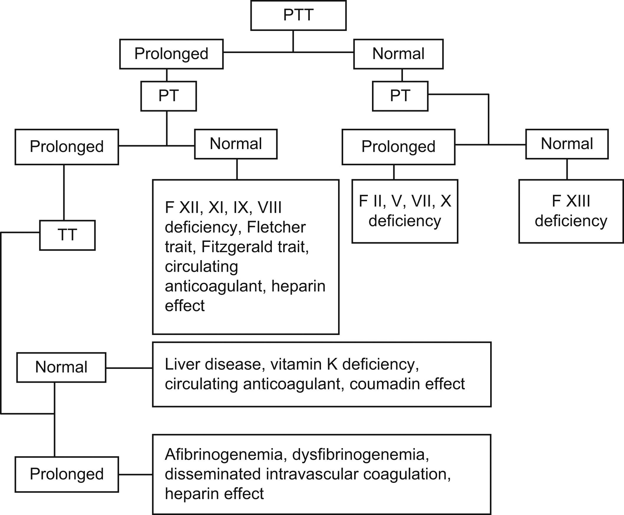
Test your understanding!
A 65-year-old patient with severe sepsis is noted to be bleeding from IV sites.
Labs show:
- PT/INR: 2.8 (Normal ~1.0)
- PTT: 75 seconds (Normal 25-40 sec)
- Platelets: 45 x 109/L (Normal 150-400)
Which pattern is present, and what is the most likely underlying cause given the clinical context?
Show answer
The pattern is a prolonged PT/INR and PTT. This points to a problem in the common pathway or a global consumption of factors. Given the clinical context of severe sepsis, bleeding, and associated thrombocytopenia, the most likely diagnosis is Disseminated Intravascular Coagulation (DIC).
Next Steps: Deficiency vs. Inhibitor
When you encounter a prolonged PTT or PT, a key follow-up question is whether the cause is a deficiency of a factor or an inhibitor (like an antibody) that is neutralizing the factor. The test to differentiate these is a mixing study.
In a mixing study, the patient's plasma is mixed 1:1 with normal plasma (which contains 100% of all factors).
- If the prolonged test corrects (becomes normal), it means the normal plasma supplied the missing factor. The diagnosis is a factor deficiency.
- If the prolonged test does not correct, it means an inhibitor in the patient's plasma is neutralizing the factors from the normal plasma as well.
This concept is briefly mentioned in the Strong Medicine video you watched (section starting at 17:43) and is the logical next step in a hematology workup.
Conclusion
You have now learned the foundational framework for interpreting screening coagulation tests. By understanding the underlying cascade and what each test measures, you can transform a pair of numbers into a powerful diagnostic tool.
Key Takeaways:
- The coagulation cascade is a map with three routes: extrinsic, intrinsic, and common.
- PT/INR is your tool to check the extrinsic + common pathways.
- PTT is your tool to check the intrinsic + common pathways.
- Isolated Prolonged PT/INR points to a Factor VII issue (e.g., early vitamin K deficiency, warfarin).
- Isolated Prolonged PTT points to an intrinsic pathway issue (e.g., hemophilia, heparin, factor inhibitors).
- Prolonged PT/INR and PTT points to a common pathway or multi-pathway problem (e.g., liver disease, DIC, severe Vitamin K deficiency).
In our next lesson, we will build directly on this foundation. We'll take a deeper dive into two of the most common clinical causes of these abnormalities: vitamin K deficiency and liver disease, and learn to correlate their specific effects with the PT/INR and PTT results.