Welcome back! In our previous lesson, we conducted a clinical review of the anterior segment and ocular adnexa, connecting structures like the cornea, lens, and drainage angle to conditions such as cataracts, dry eye, and the mechanisms of glaucoma.
Today, we move to the back of the eye and beyond. This lesson will outline the anatomy of the posterior segment and the visual pathway. Our objective is to connect the major structures—the retina, optic nerve head, and the neural pathway to the brain—with significant pathologies you regularly encounter, including age-related macular degeneration, glaucomatous optic neuropathy, and visual field defects.
We will cover:
- The anatomy of the retina and macula and their connection to age-related macular degeneration (AMD).
- The structure of the optic nerve head and its characteristic changes in glaucomatous optic neuropathy.
- The full visual pathway from the optic nerve to the brain, correlating lesions along this path with specific visual field defects.
1. The Retina, Macula, and Age-Related Macular Degeneration
The retina is the neural tissue lining the back of the eye that converts light into electrical signals. Given your background, you'll be familiar with its complex, layered structure. Let's refresh our understanding of its key components and link them directly to one of the most common retinal pathologies.
The following reading provides a concise yet thorough review of retinal anatomy and its most common conditions.
Retinal Vision Guide: Anatomy, Function & Common Conditions
This article from BrightSight, titled "Retinal Vision Guide", will serve as a great refresher on the retina's structure, function, and its link to age-related macular degeneration.
Please read the following sections: Anatomy of the Retina: Read this entire section to review the retinal layers, the specific anatomy of the macula and fovea, the role of photoreceptors, and the dual blood supply. Age-Related Macular Degeneration (AMD): Find this subsection under 'Common Retinal Conditions' and read it carefully. \nWhile reading, focus on why the macula's unique anatomical features (high concentration of cones, reliance on the RPE and choroidal blood supply) make it particularly vulnerable to the changes seen in AMD.
As the article highlights, the macula is the epicenter of our high-acuity, central vision. Its health is paramount for tasks like reading and recognizing faces. The pathology of AMD is directly tied to its unique anatomy:
-
Dry (atrophic) AMD: This form is characterized by the buildup of metabolic waste products (drusen) between the retinal pigment epithelium (RPE) and Bruch's membrane. The RPE is vital for nourishing the photoreceptors, and this disruption leads to the slow, progressive atrophy of RPE cells and the overlying photoreceptors, causing a gradual decline in central vision.
-
Wet (exudative) AMD: This more aggressive form involves the breakdown of Bruch's membrane, allowing fragile, abnormal blood vessels from the choroid to grow under the retina (choroidal neovascularization). These vessels leak fluid and blood, causing rapid and severe damage to the macula, leading to distorted vision (metamorphopsia) and central blind spots (scotomas).
2. The Optic Nerve Head and Glaucomatous Optic Neuropathy
In the previous lesson, we discussed glaucoma from the perspective of intraocular pressure (IOP) and the anterior chamber's drainage angle. Now, we'll focus on where the damage actually occurs: the optic nerve head (ONH). Glaucoma is fundamentally a progressive optic neuropathy characterized by the death of retinal ganglion cells and the thinning of their axons, which make up the optic nerve.
The following video provides an in-depth clinical look at the structural changes that occur at the optic nerve head in glaucoma. This level of detail should align well with your existing knowledge from optometry school and clinical practice.
OPTIC DISC CHANGES IN GLAUCOMA
This video, "OPTIC DISC CHANGES IN GLAUCOMA" from Insight Ophthalmology, details the specific morphological signs used to diagnose and monitor glaucomatous damage.
Please watch the following key segments: Definition of Glaucoma (00:00 - 01:33): A quick review of glaucoma as a progressive optic neuropathy. Anatomy of the Optic Nerve Head (01:56 - 05:46): Focus on the lamina cribrosa and why the superior and inferior poles are most susceptible to damage. The Neural Retinal Rim & ISNT Rule (13:52 - 16:15): A refresher on this fundamental rule for assessing rim health. Cupping and Notching (16:15 - 21:00): Understand how generalized cupping and focal notching represent nerve fiber loss. Peripapillary Changes (33:56 - 38:20): Pay close attention to the clinical significance of disc hemorrhages and retinal nerve fiber layer (RNFL) defects. \nYour focus should be on recognizing the patterns of structural damage that define glaucomatous progression.
The video provides an excellent clinical summary. The key is to connect the loss of retinal ganglion cell axons to the visible changes at the optic disc.

To summarize the clinical signs of glaucomatous optic neuropathy:
- Increased Cup-to-Disc Ratio (CDR): As axons are lost, the neural retinal rim thins, causing the central cup to enlarge. A vertically oval cup is particularly suspicious.
- Notching: Focal loss of the neural retinal rim, most commonly at the inferior and superior poles, creating a "notch."
- Retinal Nerve Fiber Layer (RNFL) Defects: These appear as dark, wedge-shaped areas on the retina, representing the loss of axons. They often correspond to an area of notching.
- Disc Hemorrhages (Drance Hemorrhages): Splinter-shaped hemorrhages at the disc margin are a strong indicator of active glaucomatous progression.
Observing these changes is how glaucoma is diagnosed and monitored, often more so than relying on IOP measurements alone.
3. The Visual Pathway and Field Defects
Finally, let's trace the journey of the visual signal from the retina to the brain and see how damage along this path creates predictable patterns of vision loss.
This animated video clearly illustrates the entire afferent visual pathway, from the optic nerve to the primary visual cortex.
CN 2: Optic Nerve (Scheme, Pathway, Lesions) | Neuroanatomy
This video from Taim Talks Med, "CN 2: Optic Nerve", provides a clear and concise overview of the visual pathway and the clinical deficits associated with lesions at different locations.
Please watch the following segments: Retinal Neurons (06:01 - 07:53): A quick recap of the first three neurons in the pathway. Visual Fields & Optic Chiasm (07:53 - 09:48): Focus on how visual fields are represented on the retina and the crucial decussation (crossing over) of nasal fibers at the chiasm. The Full Pathway (09:48 - 14:45): Follow the axons through the optic tract, the lateral geniculate nucleus (LGN), and the optic radiations to the occipital lobe. Clinical Relevance (18:37 - 21:18): This section directly connects lesions to specific visual field defects.
The organization of the visual pathway is precise, which is why the location of a lesion can often be predicted by the specific type of visual field defect a patient presents with.
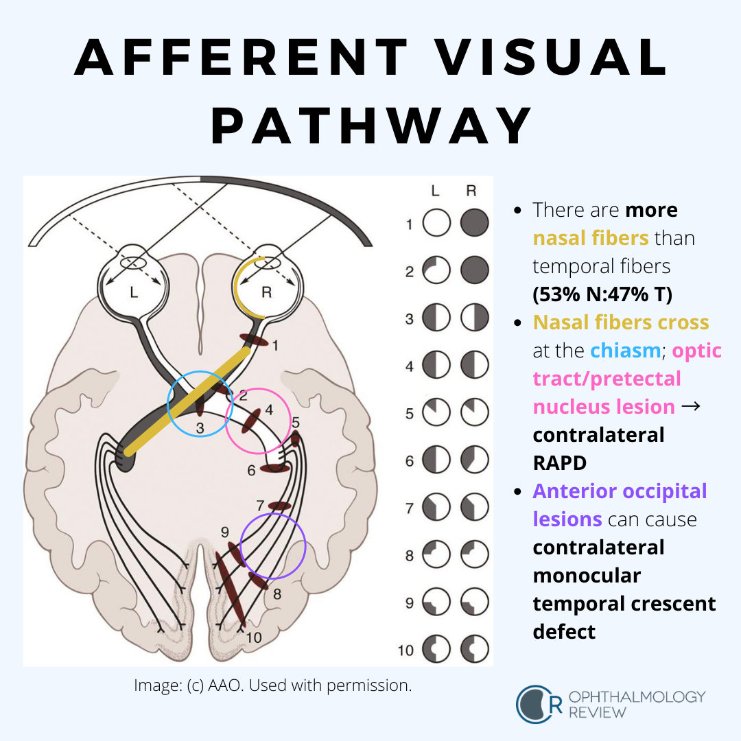
The fundamental rules for localizing lesions are:
- Lesion of the Optic Nerve (Pre-chiasmal): Causes vision loss in one eye only (e.g., complete blindness or a scotoma in the ipsilateral eye).
- Lesion at the Optic Chiasm: Typically compresses the crossing nasal fibers from both eyes, resulting in a bitemporal hemianopia (loss of the temporal visual fields in both eyes). This is the classic sign of a pituitary adenoma.
- Lesion of the Optic Tract or Optic Radiations (Post-chiasmal): Damages fibers from both eyes that represent the same half of the visual field. This causes a contralateral homonymous hemianopia (e.g., a lesion in the right optic tract causes loss of the left visual field in both eyes).
Understanding these relationships is a cornerstone of neuro-ophthalmic diagnosis.
Conclusion
In this lesson, we completed our journey through the eye by exploring the posterior segment and the visual pathway to the brain. We reinforced the anatomical basis of several major ophthalmic conditions.
Key Takeaways:
- Retina and Macula: The macula's high metabolic demand and unique structure make it susceptible to AMD, which manifests as drusen and atrophy (dry) or abnormal vessel growth (wet).
- Optic Nerve Head: Glaucoma is an optic neuropathy causing characteristic structural damage, including rim thinning, cupping, notching, and RNFL defects, which are crucial for diagnosis and management.
- Visual Pathway: The precise anatomical organization of the visual pathway means that lesions produce predictable visual field defects, allowing for accurate localization of the underlying problem.
This concludes our two-part rapid review of clinically relevant eye anatomy. You have now refreshed your knowledge of both the anterior and posterior segments, connecting key structures to the pathologies you will frequently manage in a clinical setting.